If a doctor tells you that you would need surgery – kidney tumor, prostate cancer, urinary blockage, or another urological issue – your first few questions would probably be, “Will I be out of work long?” “How big of a cut do I have to get?” “Will I need to be in the hospital overnight?” and “What is the full recovery time?” The answers to these questions are highly variable.
Laparoscopic and robotic surgeries have made the need to ask these questions virtually obsolete. Traditional surgeries of the 20th century required large cuts. Modern robotic and laparoscopic surgery techniques are conducted through cuts that are only a centimeter in length. This means that patients heal much faster after the surgery is less extensive. Robotic surgeries are able to provide the same if not better surgical or cancer control as traditional open surgery would provide.
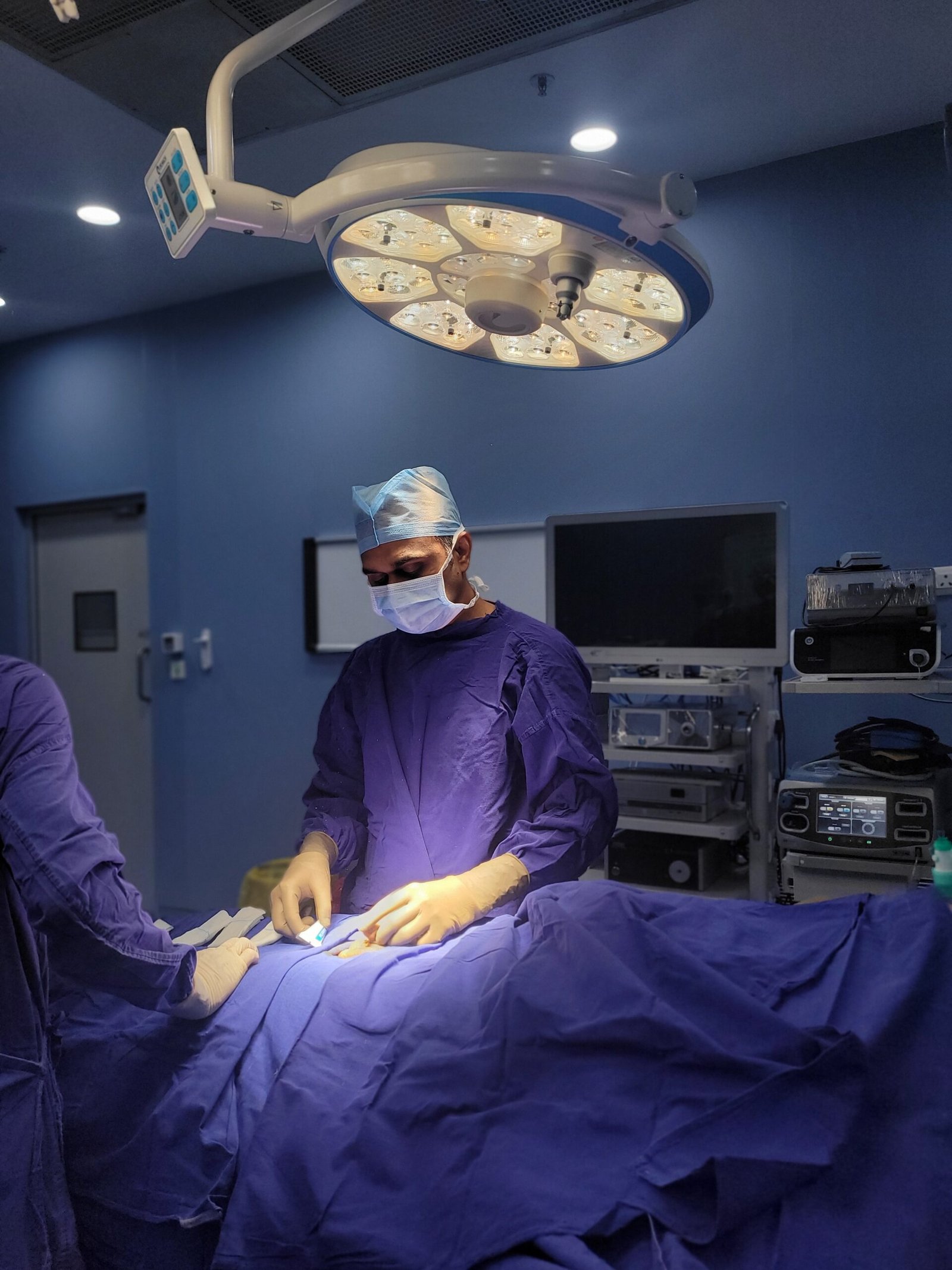
Dr. Vikas Singh, Senior Consultant Urologist at Kokilaben Dhirubhai Ambani Hospital, Nipania, Indore, is among the most skilled surgeons specializing in minimally invasive urology in Central India. Having expertise in over 15 years of performing thousands of laparoscopic and robotic urological surgeries at PGI, Chandigarh, Max Super-Speciality Hospital, New Delhi, and SMS Medical College, Jaipur, advanced robotic surgeries is at the fingertips of the people of Indore, and the entire Madhya Pradesh region, as they will no longer need to travel to the big metro cities.
In understanding laparoscopic and robotic surgery, it is necessary to first consider what open surgery entails. Open surgery requires the surgeon to make a single long incision, anywhere from 10 to 25 cm long, through the skin and muscle to obtain direct access to the organs. While open surgery is efficient, this causes a lot of trauma to the target and surrounding tissue and, as a result, requires longer to heal, longer hospital stays, more post-operative pain, and longer recovery times to return to baseline activities.
Laparoscopic surgery changes the status quo. Rather than having a single long incision, multiple insertions (3 to 5) of cuts that measure anywhere from 5mm to 12mm (about the size of a fingertip) is the standardized practice. The small cuts (port holes) are the entry point of a slick, high-definition camera (with an elongated apparatus that is surgical) with the cut measuring 5mm. This small 5mm port, after an incision with a variety of high-definition apparatuses) becomes an (effectively 5mm) open window to the body, such that the surgeon is able to get an incredibly close (real-time) view of the inner body, and is a direct means of surgical control.
To image the body cavity, the abdomen is deliberately inflated with CO2 at the start of the surgery and is eventually absorbed at the end. The use of CO2 is a measure taken in the absence of an in-body control space to facilitate operative control.
An advancement on traditional laparoscopic surgery is robotic surgery. A robotic surgical system, like the da Vinci Surgical System, is placed over the patient. Rather than controlling the robotic arms and surgical instruments at the surgery table as he or she would for laparoscopic surgery, the surgeon commands the arms from a console. The robotic arms are able to fill in any hand twitch, let the surgeon control the arms at a range that human hands are not able to, and replicate the movements of the surgeon. The modular arms are able to weave through the body more easily than traditional laparoscopic surgery instruments.
As a result of these factors, robotic surgery is comparable to open surgery for the surgeon’s hand in regard to control over body functions and for cancer-related surgery. However, robotic surgery is less severe to the body, involves less blood loss and less pain, and is associated with shorter hospital stays and a quicker recovery.
It is important to understand the differences between robotic and laparoscopic surgery when discussing the details about a patient’s condition with Dr. Vikas Singh, as both techniques are minimally invasive and use a camera and small incisions.
The surgeon using the keyhole method controls the surgery from the operating table. The surgeon uses long, thin instruments and controls the surgery with his or her own hands. The surgery is viewed on a monitor, which may show a 2D or 3D image depending on the scope used.
As laprascopic techniques have evolved over the past 30 years, the surgery techniques have been simplified. Procedures such as donor nephrectomy, removal of a kidney, or pyeloplasty that once required large incisions of 15-25 cm can now be performed through 3-4 small incisions and still be oncologically and functionally sound. The recovery timeframe has decreased from 4-6 weeks to 1-3 weeks.
Kokilaben Hospital’s Dr. Vikas Singh has performed many rarius laparoscopic surgery techniques on his patients. His specialties include, but are not limited to, laparoscopic nephrectomy (partial and radical), laparoscopic donor nephrectomy, and pyeloplasty, as well as, laparoscopic vesicovaginal repair.
Robotic surgery enhances the precision and effects of laparoscopic surgery, providing:
Three-dimensional (3D) enhanced, high-definition vision. 3D camera depth is superior to 2D camera depth. Surgeons have the ability to judge the distance of tissue planes and structures.
Wristed instrument movement. Laparoscopic instruments are limited to a few degrees. Surgical instruments utilize a wrist to spin instrument tips and achieve 360-degree movement. Deep to the pelvis injuries will require substantial manipulation.
Robotic surgery eliminates errors due to the imperfections of the hand that the surgeon is manipulating. Tremors, shakes, and natural imperfections will be eliminated.
Robotics provides a 10 to 15 times magnification and improvement that is significant to standard laparoscopy. Precise structure identification (nerves, vascular elements and other structures) is improved considerably.
Robotic surgery is superior to standard open surgery for injuries deep to the pelvis for (prostate) injuries due to the impact of the face touch system control, superior vision, and elimination of tremors that provide substantial improvement in patient cancer control and improvement in the loss of sexual and urinary control. Robotic surgery is the gold standard for the improvement of the treatment of multiple urological disorders. Customers.
Advantages and disadvantages of the three surgical techniques in the same urological procedure:
Factor | Open Surgery | Laparoscopic Surgery | Robotic Surgery |
Incision Size | 10–25 cm (large single cut) | 3–5 ports of 5–12 mm each | 3–5 ports of 5–12 mm each |
Blood Loss | Higher – larger dissection | Significantly less | Least – finest dissection |
Post-op Pain | Significant – larger wound | Mild to moderate | Mild – minimal tissue trauma |
Hospital Stay | 5–10 days typically | 2–4 days typically | 1–3 days typically |
Recovery Time | 4–8 weeks | 2–4 weeks | 1–3 weeks |
Precision | Good with direct vision | Very good with camera view | Excellent – 10–15x magnification, 3D, wristed instruments |
Nerve Sparing | Possible but limited by view | Better visualisation | Best – superior nerve identification |
Infection Risk | Higher – larger wound | Low | Very low |
Cosmetic Result | Large visible scar | Tiny port scars | Tiny port scars |
Best For | Very large tumours; emergency surgery; selected complex cases | Most standard urological procedures | Complex pelvic surgery; nerve-sparing; kidney-sparing tumour surgery |
While modern methods are important, the recommended surgical approach must be based on the patient’s unique condition, anatomy, and the judgment of the surgeon. There are cases where open surgeries can be the best applicable method. Dr. Vikas Singh advocates for his patients adopting a similar approach.
Dr. Vikas Singh uses the latest technology for laparoscopic and robotic surgery for all urological cases at Kokilaben Dhirubhai Ambani Hospital, Indore. The following are briefly described common surgical cases:
A partial nephrectomy is the excision of a kidney tumor with removal of a minimal amount of surrounding normal renal tissue. This approach is highly preferred over the radical nephrectomy (i.e. removal of the entire kidney and all surrounding tissue) for tumors located in the kidney that are smaller than 7 cm and do not involve the renal vessel, in order to preserve the patient’s remaining renal function. This is of utmost importance for individuals with diabetes, hypertension, or those who have a single kidney left.
Advances in laparoscopic and robotic techniques have vastly improved the ability to perform robotic and laparoscopic partial nephrectomy versus open partial nephrectomy. The robotic systems’ ability to obtain a highly magnified view of the surgical field allows Dr. Vikas Singh to differentiate normal renal tissue from the tumor and the surgical field, achieve excellent oncologic margins, and suture the remaining normal renal parenchyma, all through small incisions. Additionally, the warm ischaemia time is the period during which blood flow is interrupted, is generally prolonged in open surgical approaches versus robotic laparoscopic approaches, and is especially minimized because of the nature of robotic surgery.
Most patients are discharged within 2 to 3 days, compared to 5 to 7 days for open partial nephrectomy. Patients can also return to their normal routines within 2 to 4 weeks. Laparoscopic and robotic techniques for nephrectomy also have comparable long-term oncological outcomes, and nephron-sparing surgery is superior to complete nephrectomy for the preservation of renal function.
The goal of a radical prostatectomy is a total surgical removal of the entire prostate gland as well as the nearby tissue and the seminal vesicles. Localized prostate cancer is surgically remedied in this manner and there is no method of treatment that is more certain. Although there are many surgical procedures for which the value of robotic versus laparoscopic surgery is evident, radical prostatectomy is one of the more obvious cases. 25
The prostate is an organ that is located internally within the male’s pelvic cavity. Structures that are positioned around the prostate and the surrounding tissue that are of critical importance and provide a quality of life for the individual include the urinary sphincter.; This is the muscular device that controls urethral continence; the neurovascular bundles that flank the side of the prostate which provide the support for erectile function; and the neck of the bladder located above the prostate.
Direct access, let alone intimate access, to the surgical field using the old school techniques is not practical. Scarce surgical field access precludes nerve saving procedures from being performed.
Advancements in the area of laparoscopic/robotic radical prostatectomy have resulted in the development of instruments that provide the surgeon a 3D, magnified view of the surgical field as well as instruments that allow for the same flexibility as the surgeon’s wrist. This allows the surgeon, in this case Dr. Vikas Singh, to operate in the deeper parts of the pelvic cavity with a level of precision that previously could not be achieved.
As a result of these advancements, nerve saving procedures have resulted in the protection and preservation of the neurovascular bundles. This, in turn, results in a quicker return of urinary continence and erectile function. These aspects are some of the top priorities for the patient after surgery.
At Kokilaben Hospital, after a robotic or laparoscopic radical prostatectomy, the average length of hospital stay is 2-3 days. The foley catheter is typically removed at a week. Most patients are back to regular daily activities in 3-4 weeks post surgery. A typical open prostatectomy typically results in a length of hospital stay of 5-7 days. The foley catheter is usually left in place for an extended time. The recovery time is generally 6-8 weeks.
VVF refers to a tunnel-like passage that develops between the bladder and the vagina that causes the vaginal environment to remain wet due to the constant and uncontrollable passage of urine through the duly formed channel. The presence of VVF can be the most detesting Urogynaecological condition. It can lead to the suffering person’s social isolation due to the constant wetness of the lower half of her body, odor and severe skin complication.
In India, the most common causes of the obstetric, gynaecological and fistula (VVF) causes of fistula are prolonged obstructed labour, the practice of instrumental vaginal delivery of babies, Caesarean section, and the practice of hysterectomy and/or radiation for gynaecological cancer. In India the social stigma of a constant urine leak often forces a woman to live with her condition (s) for years in relative embarrassment and in complete isolation.
The use of laparoscopic techniques with the aid of advanced technology provides a new hope for even better and more successful outcomes. Dr. Vikas Singh will introduce small ^11-12 mm keyhole ports to the abdominal wall through which he will be able to identify the fistula tract and will be able to excise and meticulously close the fistula. Closure will be done in such a manner that the vaginal and bladder walls will be closed in a watertight fashion with absolutely fine vascular suture. As an advanced modern laparoscopic closure technology, the use of closure of the fistula will provide the scope of complete closure of the fistula. Dr. Vikas Singh uses three to four of this technology to achieve the scope of closure.
There are several advantages of laparoscopic over the traditional open approach. These benefits include a much lower risk of a hysterectomy, much less expected blood loss and a shorter expected postoperative stay ( 3-5 days vs 7-10 days). Laparoscopic implementation of a vaginal fistula closure (VVF) offers many advantages. As a result of the expected return to normal activities, many women who have undergone this procedure have discovered a new and unexpected lease on life. The recurrence rate of fistula and the expected long-term outcomes also improve in the hands of an experienced laparoscopic surgeon. multiple laparoscopic ports will be employed to provide Dr. Vikas Singh with the most advanced modern technology available for laparoscopic surgery
Pyeloplasty is done to correct the surgical problem of ureteropelvic junction (UPJ) blockage. Ureteropelvic junction (UPJ) is the junction of the funnel-shaped (pelvic) collecting part of the kidney and renal tubule part (ureter) which collecting gathers and carries the urine to the bladder. Ureteropelvic junction obstruction (UPJ) will result in urine being unable to exit the kidney. This causes collection of urine in the kidney resulting in swelling of the kidney (hydronephrosis) and eventually leads to progressive kidney damage and severe infections.
UPJ obstruction can either be congenital having been from birth (often detected during an ultrasound during pregnancy) or in childhood or acquired which occurs during adulthood (due to, for instance, a crossing blood vessel that presses on the Junction to the UPJ). Symptoms of UPJ obstruction can include flank pain that occurs intermittently, is usually heightened after the consumption large volumes of liquids; recurrent urinary tract infections (UTI’s), hematuria (blood in urine), or the presence of a kidney observed to be hydronephrotic as found incidentally.
Of the several options available for UPJ obstruction, laparoscopic pyeloplasty is the default choice of Anderson-Hynes dismembered pyeloplasty and has also overshadowed traditional open pyeloplasty in favor available in most centers. It involves excising the narrowed portion of the UPJ segment, reconstructing the renal pelvis and then reattaching the ureter in a wide funnel-shaped configuration. This restores proper drainage of urine from the kidney. During the surgery, a temporary internal ureteric (DJ stent) is inserted to prevent damage to the repair as the urine will be draining freely. This stent is removed after 4 to 6 weeks.
With intervals of lesser pain, shorter stays of just 2 to 3 days, and quicker return to normal activities within 2 to 3 weeks, Laparoscopic pyeloplasty at Kokilaben Hospital provides patients with an equal success rate of 90 to 95%, as that of an open pyeloplasty case.
The process of Laparoscopic donor nephrectomy, is considered to be the most patient-centric procedure within all of urology, as it involves the removal of a donor’s kidney for the purpose of the donor’s kidney removal for the purpose of the life salvation of the donor’s. Naturally, it is an ethical and medical obligation that surgery is as minimally disruptive to the donor as possible in terms of pain, recovery time, leave from work, and social engagement.
This is Laparoscopic donor nephrectomy. Dr. Vikas Singh makes 3 to 4 incisions, through which he painstakingly removes the donor’s kidney. In doing this, he preserves the renal vessels and ureter to the maximum length. This is so as to technicaliazz the success of the transplant. The kidney is removed at the end of the procedure through a small extension of one of the small incisions. The incision is ranged to be between 4 to 5 cm.
Donor experiences during laparoscopic nephrectomies are generally better compared to open nephrectomies. Laparoscopic procedures typically result in substantially less pain and leave behind smaller scars that are easier to hide. Hospital stays are shorter, averaging just 2-3 days compared to 5-7 days, and a nephrectomy patient can return to work more quickly, in just 2-3 weeks instead of 4-6 weeks. The outcomes of the grafts are also comparably successful.
These reasons, among others, encourage more people to donate kidneys. They know with limited lifestyle and health impacts, they can help a patient in need. Dr. Vikas Singh has completed many laparoscopic donor nephrectomies and has gained experience in the field of transplantation.
The importance of using minimally invasive surgical techniques compared to traditional open surgeries that require long skin incisions goes beyond one simple reason. The benefits are large both in number and in their clinical importance.
When compared to traditional open surgical techniques, minimally invasive approaches lead to smaller incisions, ranging from 1-3 surgical portal incisions of diameters 5mm to 12mm, compared to the large 10-25cm incisions associated with traditional open surgeries. This is because incision size is proportional to the size of the complete incision needed to access the surgical site. Minimally invasive approaches, when compared to traditional open surgeries, are associated with less perioperative blood loss.
Open surgeries are associated with high risks of perioperative blood loss, frequent blood transfusions, postoperative anemia, and high strain on the cardiovascular system. Robotic and laparoscopic surgical approaches are associated with high control, lower risks of cutting large blood vessels, and high risks of high blood loss, confirming the benefits of both approaches.
The stress placed on the supportive tissues (muscles, etc.) is less when compared to traditional open approaches, and aids in faster recovery.1 Most laparoscopic surgeries lead to a postoperative hospital stay of 1-3 days, compared to 5-10 days for open surgeries.
Unlike open surgery where recovery can take 4 to 8 weeks, most patients can resume their light activities in less than 2 weeks, and normal activities in less than 4 weeks. This represents less time off work, less reliance on help from family and friends during the recovery process, and a faster return to daily life.
Post-operative pain after surgery is often proportional to the degree of tissue injury involved. Because no large incision with healing for the patient is involved in laparoscopic and robotic surgery, the patients often need mild to moderate pain medication for only a few days. The recovery is frequently surprising, and they often feel comfortable after what they expected to be a much more painful.
An even greater risk of large, open incisions is the risk of a wound infection. Because there is a large reduction in the potential for wound contamination in keyhole surgery, there is a reduction of the wound surface as well. Port site infections can occur, but are rare, minor, and easily managed. In experienced hands, laparoscopic and robotic techniques greatly reduce the risk of deep surgical infections.
Surgeons now have the ability to see the anatomy in far greater detail thanks to the modern laparoscopes and robotic systems, which provide a magnified, high-definition view. This technology enables surgeons to detect nerves and blood vessels and tissue planes in a way that exceeds the natural visibility of open incisions. This heightens the overall surgical accuracy and ultimately results in better control of cancer margins, higher quality and preservation of nerved tissue in prostate cancer surgery, as well as improved results with reconstructive surgeries.
Robotic surgery’s ability to magnify images tenfold to fifteenfold along with the introduction of three-dimensional imaging and the ability of an instrument to reflect the arthritic-like movements of a surgeon makes robotic surgery devices superior for some of the more complex surgeries including surgeries for pelvic and retroperitoneal structures. The ability of the systems to filter a surgeon’s hand tremors allows the device to facilitate deliberate and steady movement. For example, the device can be used to carefully dissect a nerve bundle from a tumor and to take a suture from a 3 mm blood vessel.
Being familiar with the surgical journey can reduce anxiety about the process. Here is a straightforward guide about the laparoscopic and robotic surgeries conducted by Dr. Vikas Singh at Kokilaben Hospital, Indore, to construct a vision of the process.
Before any laparoscopic or robotic surgeries, a thorough evaluation has to be completed. This includes:
You arrive days before the surgery is scheduled to begin. The operating room staff prepares for IV placement and hemodynamic monitoring before the start of general anesthesia.
Once under anaesthesia and positioned on the operating table, the surgical team prepares the keyhole incisions. The abdomen is gently inflated using CO2 through a small needle. The incisions for the camera and surgical instruments are also prepared.
In robotic surgery, arm appendages of the robot are positioned and linked to the instrument ports. Robotic surgery is done from a console. A surgical assistant standing next to the table makes the instrument exchanges and tablets, and supports further the console.
This procedure is done intra-operatively with high definition imaging for an enlarged view of the field. Robotic systems and instruments are used to remove a tumor, control bleeding, and for other complicated tasks, The procedure lasts 1 to 4 hours
To finish the procedure, the instruments are taken out and the camera ports are taken out. The abdomen is no longer inflated, and the incison ports are closed. The ports are covered with a sterile dressing. A urinary catheter covered with sterile dressing is also done. The catheter remains for 1 to 2 weeks.
Most people wake up from general anesthesia in a recovery room and spend 1 to 2 hours getting monitored before being moved to a ward. Nausea from anesthesia is very common and passes within hours of waking. After 24 to 48 hours of surgery, gas from the procedure may make your shoulder and/or upper abdomen hurt, but this too passes and is normal.
Below is what you can expect from your recovery in the days and weeks after having robotic or laparoscopic surgery.
Days 1 to 2
Your recovery begins in the hospital. You will receive IV fluids and meds while your progress is monitored. You will be asked to walk and move around to promote recovery, as well as to reduce the risk of blood clots. Your recovery will be helped further by a clear fluid diet that progresses to light solids. Meds will be given to you to manage your pain and most cathaters will be taken out in under 24 hours; however, this is not the case with prostate surgery, where the cathater will remain for an entire week.
Days 3 to 7
You will no longer be monitored, but recovery will still be an active process. You will be responsible for your own recovery and will be instructed on how to proceed. Recovery wounds will be small, and will need to be protected in a clean and dry manner. You cannot drive for 5 to 7 days. For 2 to 3 weeks, do not lift anything that weighs more than 5 kg. For 2 to 3 weeks, you will need a balanced light diet. For recovery passes, you will receive medications including antibiotics. You will also receive pain medications and blood thinners. You must take each of these as directed.
Weeks 2 to 4
Gradual Recovery: Most patients return to light desk work in about 1 to 2 weeks. Driving can start in the same timeframe. Moderate exercise including walking and light stretching may begin at 2 weeks and swimming, gym workouts, and other strenuous activities can begin at 3-4 weeks (or as recommended by Dr. Vikas Singh based on the specific procedure.)

Dr. Vikas Singh is one of Central India’s top laparoscopic and robotic surgeons. Due to his performance of thousands of laparoscopic and robotic urological procedures such as nephrectomy, pyeloplasty, prostatectomy, VVF repair and donor nephrectomy, he has been able to perfect and hone his skills as one of the very best urological surgeons in the field. He has now brought his craft to Indore so those living there do not have to travel anywhere to receive highly skilled surgical practice. This depth of volume and experience is certainly rare in Central India.
In the case of laparoscopic surgery, many medical professionals view it as equally safe, if not more so, than traditional open surgery. Traditional open surgery involves making more big cuts, leading to possible loss of blood, as well as the risk of infection, and the formation of blood clots. When performed by a surgeon as skilled as Dr. Vikas Singh, the outcomes in terms of keeping the cancer under control, as well as the symptoms of that cancer, such as loss of urinary continence after surgery, will be as good if not better than traditional open surgery.
There are very few patients who will present symptoms that rule them out of the robotic and laparoscopic surgery pathway. There are a few conditions that will affect a person’s eligibility. If a person has extremely high body mass, has undergone several large abdominal surgeries, has a cardiovascular condition, or has a large tumor that requires a high level of dissection, such a person may be ineligible for robotic surgery or laparoscopic surgery. Dr. Vikas Singh will review your case and determine the level of surgery that will be most appropriate for you.
Not in the case of laparoscopic or robotic surgery. There are two types of surgeries that may be involved – laparoscopic and robotic surgery. Either type of surgery will be performed under general anesthesia. Therefore, you should feel nothing at all. The anesthesia team will take care of everything and monitor your condition even after the anesthesia has worn off and the surgery is a thing of the past.
Vikas Singh says hospital stay post surgery varies depending on the specific procedure. 2-3 days is the typical hospital stay after nephrectomy, pyeloplasty, and donor nephrectomy. A 3 to 5 day hospital stay is typical post laparoscopic VVF repair. Laparoscopic radical prostectomy usually requires hospital stay of 2 to 4 days. A longer catheter stay at home is typical.
Participants that have port scars due to laparoscopic or robotic surgery will usually notice that the scars become less noticeable in as short a time scale as between 2-12 months. Many of these scars occur in the lower abdomen or around the abdominal cavity as they range from 5-12mm in size.
Flat, 2D cameras provide no perception of depth while 3D laparoscopic systems use special cameras to offer depth perception. This allows the surgeon to judge distances and tissue planes more accurately. High-definition, 3D robotic vision is the standard. At Kokilaben Hospital, Dr. Vikas Singh uses advanced laparoscopic systems with 3D and 4K imaging coupled with the robotic platform for selected procedures.
The need for a blood transfusion after laparoscopic or robotic surgery is much less likely than after open surgery. This is due to precision of laparoscopic or robotic surgery and the reduction of blood loss. There is a complication with every procedure and unexplained or unexpected issues with bleeding could necessitate a blood transfusion. Dr. Vikas Singh’s team ensures that cross-matched blood is readily available for every major surgery undertaken.
The carbon dioxide gas that is used to inflate the abdomen deflates and releases the gas at the end of the surgery at the time of closure of the laparoscopic ports. Any residual carbon dioxide in the abdomen is absorbed in a matter of a few hours and exhaled by the lungs in a day or two. This is the cause of the minor pain and discomfort in the shoulder or upper abdomen after surgery for a day or two and is due to the residual gas irritating the diaphragm. This is self limiting and is relieved with ambulation and movement.
Yes – laparoscopic pyeloplasty (Anderson-Hynes dismembered pyeloplasty) performed at leading centres, has success rates for permanent renal blockage cure approaching 90-95%. After surgery, patients typically do not experience further kidney pain, pyelonephritis, or hydroureter/hydronephrosis. To facilitate kidney healing, a ureteral DJ stent will be placed and later removed 4-6 weeks postoperatively. Post-surgery, a MAG3 renal scan will be conducted at 3 months to confirm successful kidney healing.
24/7 Services Available
Copyright © 2026 Urology Center | All Rights Reserved
Design and Developed by Namastetu Technologies Pvt.Ltd