Male infertility is much more common than most people realize, it makes up some 40-50% of all infertility cases. And varicocele, the abnormal dilation of veins draining the testicle, is the most common of the identifiable, treatable causes of male infertility. Varicoceles occur in approximately 15% of all men and up to 40% of men who present for evaluation of infertility. More importantly it is eminently treatable.
Microsurgical varicocelectomy is the gold standard for the surgical treatment of varicocele. Using high powered surgical magnification, all of the dilated internal spermatic veins are identified and ligated, while the testicular artery, lymphatic channels and the vas deferens are carefully preserved. The microscopic technique has the highest success rates, the lowest recurrence rates and the fewest complications compared to open or laparoscopic . It is the treatment of choice in experienced urology centres worldwide.
The microscope used in microscopic varicocelectomy magnifies 6-25 times and enables the surgeon to identify easily and to ligate individually all dilated veins of the spermatic cord while at the same time preserving the small testicular artery, lymphatic vessels and vas deferens. Such a level of precision cannot be achieved with the naked eye or the usual magnification of a laparoscopic camera, and the microsurgical technique has therefore been shown to give significantly better results than any other approach to varicocelectomy.
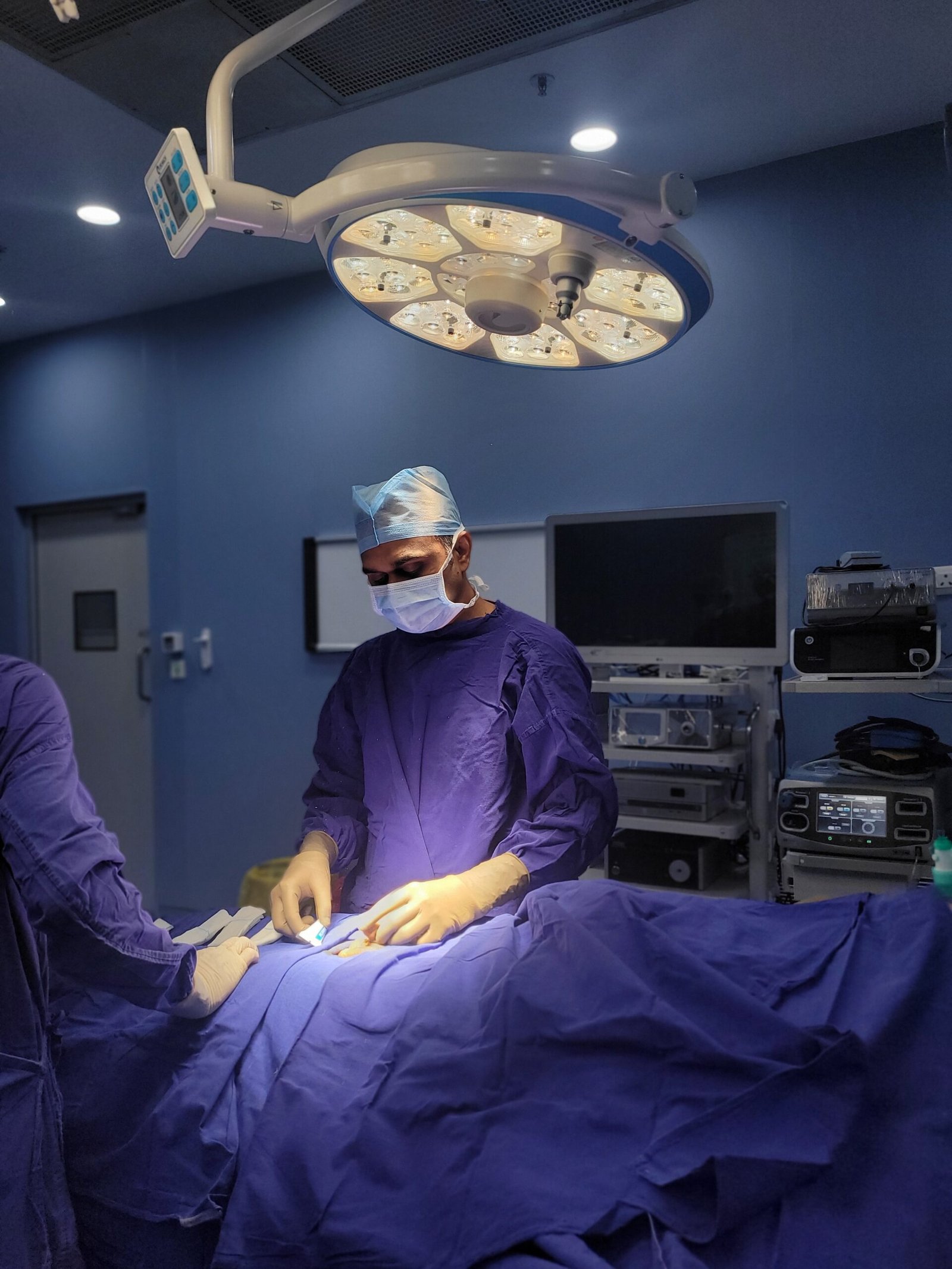
The procedure is performed through a small incision, usually 2 to 3 centimetres, at the level of the inguinal canal (groin) or at the subinguinal level, just below the external inguinal ring. The spermatic cord is delivered out through this incision and examined under the operating microscope. The surgeon then identifies each dilated vein within the cord in a systematic fashion . There are usually between five and twenty of these , once all the internal spermatic vein branches are counted . Each is ligated ( tied off ) individually . The testicular artery, a vessel less than 0.5 mm in diameter, and the lymphatic channels are identified and preserved under magnification. The entire procedure usually takes about 60 to 90 minutes a side.
This technique was pioneered in the 1990s by urologists Marc Goldstein and Peter Schlegel at Cornell University and is now the method of choice for varicocele repair in fertility centres worldwide. The magnification of the operating microscope transforms the spermatic cord from a confusing jumble of indistinguishable structures into a clearly mapped anatomy where each vessel type can be confidently identified and the appropriate surgical decision made for each one. This precision is what makes microscopic varicocelectomy so much more effective and so much safer than its nonmicroscopic alternatives.
Varicocele is an abnormal distention (varicosity) of the pampiniform plexus which is the network of veins that drains blood from the testicle and makes up the main venous drainage of the spermatic cord. Varicoceles are similar to varicose veins in the leg and develop through the same mechanism. This occurs when venous valves fail to function properly and cause a pooling of blood and retrograde flow in the network of veins rather than an efficient flow toward the heart.
The left testicular vein enters the left renal vein at a right angle leading to increased venous pressure in the left pampiniform plexus compared to the right where the testicular vein drains more directly into the inferior vena cava at a more favourable angle. This anatomical difference explains the fact that the vast majority of varicoceles, about 90 percent, are left-sided. Right sided varicoceles are much rarer. When they do occur they can sometimes suggest an underlying abdominal or retroperitoneal cause which must be ruled out with imaging (e.g. a renal or retroperitoneal tumour).
Varicoceles are classified clinically from Grade 1 (only palpable during Valsalva manoeuvre , bearing down) to Grade 3 (visible through the scrotal skin without any manoeuvre, classically described as a ‘bag of worms’ appearance). Grade 2 varicoceles are palpable on clinical examination without Valsalva manoeuvre. Subclinical varicoceles are not palpable on clinical examination but can be detected on Doppler ultrasound and their clinical significance is uncertain; they are not usually treated.
Varicocele impacts on testicular function by multiple and synergistic mechanisms. The dilated veins allow retrograde blood flow , which increases the temperature in the scrotum . The testicle normally functions at 2 to 3 degrees Celsius below core body temperature , and this thermoregulatory advantage is lost when hot venous blood pools around it . Increased scrotal temperature directly impairs spermatogenesis (sperm production), damages sperm DNA and decreases testosterone production by Leydig cells. Venous stasis also results in the accumulation of toxic metabolites (e.g ., adrenal steroids, catecholamines, and reactive oxygen species) that reflux down from the adrenal and renal veins into the testicular venous system , exacerbating the damage to testicular function. The net result of these mechanisms is progressive deterioration of semen quality and testicular volume with time.
Most men with a varicocele have no symptoms at all. The condition is usually found by accident during a fertility workup or a routine physical exam. Others have a variety of symptoms that seriously impact quality of life. If these symptoms are identified and evaluated, timely treatment can restore fertility and prevent progressive testicular damage.
Dull Aching Pain or Heaviness in the Scrotum
The most common symptom of varicocele is usually a dull aching discomfort or a feeling of heaviness in the scrotum, most often on the left side. The pain is usually described as a deep dragging ache, mild when at rest but noticeable after standing for a long time, after physical activity or at the end of the day. It is usually not severe or sharp, and a really severe acute pain in the scrotum is more likely to be a different condition (torsion, epididymo-orchitis) and should prompt immediate emergency assessment. Men often ignore the chronic, low-grade ache of varicocele, dismissing it as “normal” for months or years before seeking evaluation.
Visible or Palpable Enlarged Veins in the Scrotum
Grade 2 and Grade 3 varicoceles are dilated veins that are visible or palpable on the left side of the scrotum, particularly in the standing position when venous pressure is at its peak. The classic clinical description is a ‘bag of worms’ , a discrete tangle of enlarged, tortuous veins above and behind the left testicle which are visualized and palpable through the scrotal skin. Men often notice this abnormality themselves in the shower or when dressing and are alarmed by its appearance. The veins are soft, nontender, and collapse when the patient is supine (there is no hydrostatic pressure to drive venous congestion in the supine position).
Worsening Pain After Prolonged Standing or Physical Activity
The pain of varicocele has a characteristic positional and activity-related quality that differentiates it from other scrotal pathologies. It is always worse after long periods of standing, heavy physical exertion or vigorous activity. These activities increase intra-abdominal pressure which increases hydrostatic pressure in the dilated veins and exacerbates venous congestion around the testicle. Men in standing occupations, regular exercisers or those doing heavy physical labour often find their varicocele pain most acute at the end of a physically demanding day. Lying down relieves the pressure and the pain usually subsides within minutes.
Testicular Atrophy (Shrinking of Testicle)
Testicular atrophy, reduction in testicular volume, is one of the most important clinical findings in varicocele and its presence in an adolescent or young adult is one of the strongest indications for surgical correction. In patients with varicocele the left testicle is often smaller than the right by measurable amounts . The chronic elevation of scrotal temperature and accumulation of toxic metabolites slowly destroys the testicular tissue’s ability to produce sperm and hormones . If testicular atrophy is detected in a teenager (e.g. progressive loss of volume on serial orchidometry or testicular ultrasound measurements), this suggests that the varicocele is causing progressive damage and that early surgical correction provides the best opportunity for volume recovery and preserved fertility.
Male Infertility & Low Sperm Count Due to Varicocele
Varicocele is the most common surgically correctable and identifiable cause of male infertility. Its characteristic and consistent effect is the common occurrence of oligospermia, asthenospermia and teratospermia, alone or in combination (the so-called ‘stress pattern’ on semen analysis). Men with varicocele also have significantly higher levels of elevated sperm DNA fragmentation (damage to the genetic material carried by sperm). In infertile men with varicocele undergoing microsurgical repair, there are significant improvements in semen parameters in 60 to 80 percent of cases, and 30 to 50 percent of couples will conceive naturally within one to two years postoperatively.
Hormonal Imbalance & Low Testosterone Due to Varicocele
Varicocele impairs not only spermatogenesis but also testosterone production by Leydig cells of the testicle. In men with varicocele, serum testosterone is significantly lower than in matched controls, and the extent of testosterone impairment is related to varicocele grade and duration . Some men with varicocele may have symptomatic hypogonadism with symptoms such as decreased libido, fatigue, loss of muscle mass, mood changes and erectile dysfunction. Importantly, published studies have demonstrated that microsurgical varicocelectomy significantly increases post-operative serum testosterone levels , thus making it not only a fertility treatment but a potential treatment for varicocele-related hypogonadism in selected patients.
An understanding of the biological mechanism by which varicocele impairs fertility helps the patient to appreciate the importance of treatment and the expected improvements after surgery.
Mechanism | How It Damages Sperm & Testis | Reversibility After Surgery |
Elevated scrotal temperature | Impairs spermatogenesis; kills developing sperm cells; reduces sperm motility | Yes , temperature normalises within weeks of surgery |
Venous stasis & toxic reflux | Adrenal steroids and catecholamines from kidney/adrenal veins damage testicular cells | Yes , eliminated when reflux veins are ligated |
Reactive oxygen species (ROS) | Oxidative stress damages sperm DNA, membrane, and mitochondria | Yes , ROS levels fall significantly post-varicocelectomy |
Sperm DNA fragmentation | Damaged genetic material impairs embryo development even when sperm count is normal | Partial , DFI improves significantly after surgery |
Leydig cell dysfunction | Reduces testosterone production; causes hypogonadism | Yes , testosterone rises significantly in most patients |
Impaired blood-testis barrier | Allows auto-antibody formation against sperm (anti-sperm antibodies) | Partial , antibody levels may decrease over time post-surgery |
Progressive testicular atrophy | Loss of testicular volume over time if untreated | Yes (partial) , volume recovery especially in adolescents and young men |
The main clinical point of this table is that most of the damage caused by varicocele is reversible, because it is caused by ongoing physiologic insults (heat, toxic metabolites, oxidative stress) that are eliminated when the dilated veins are surgically ligated. This is why varicocelectomy, when done correctly and on the right patients, brings about such consistently good improvement in semen quality, and why waiting to treat it leads to cumulative damage that is more difficult to correct.
Sperm takes about 72 to 90 days to mature (spermatogenesis cycle) . This is because the improvement in semen parameters after varicocelectomy usually takes 3 to 6 months to develop as the improved testicular environment supports a new cohort of sperm through the entire process of maturation. Therefore, couples should be advised that 3-6 months should be waited before the repeat semen analysis after surgery and not be discouraged if early post-operative semen analyses show limited improvement.
Not all men with a varicocele require surgery and proper patient selection is critical to ensure that those men who will benefit the most are treated promptly. The indications below describe the appropriate surgical candidates according to the current international guidelines (American Urological Association, European Association of Urology).
Men With Varicocele & Abnormal Semen Analysis
The primary indication for varicocelectomy in the infertility setting is the presence of a clinically palpable varicocele (Grade 1 to 3) along with at least one abnormal semen analysis parameter on two separate analyses (oligospermia, asthenospermia, or teratospermia). The abnormal semen analysis confirms that the varicocele has a quantifiable effect on sperm production. The palpable varicocele confirms a surgically treatable anatomical target. The best-supported indication for microsurgical repair and the best data on improvement in fertility after surgery is the combination of clinically detectable varicocele and abnormal semen analysis in an infertile couple.
Couples With Unexplained Infertility & Male Factor Diagnosis
Some couples investigated for infertility are given a diagnosis of ‘unexplained infertility’ , where no single cause is obvious. In couples with a clinical palpable varicocele and borderline semen analysis rather than a frankly abnormal one, varicocelectomy is a reasonable therapeutic option. Borderline semen parameters that appear ‘normal’ on standard analysis may mask significant sperm DNA fragmentation or other functional deficiencies, to which the varicocele is contributing. The measurement of sperm DNA fragmentation (DFI) is increasingly used in the workup of unexplained infertility and the presence of an elevated DFI with a palpable varicocele is an additional indication for surgery.
Men With Testicular Pain & Discomfort Due to Varicocele
Varicocelectomy is indicated for symptomatic varicocele , a palpable varicocele that causes chronic scrotal pain or discomfort that significantly impacts quality of life , even in men who are not currently trying to conceive . The indication for surgery for pain is independent of the indication for fertility. After other causes of scrotal pain have been excluded and the varicocele is confirmed by clinical and Doppler ultrasound examination, microsurgical repair reliably resolves or significantly improves varicocele-associated pain in about 80 to 90 percent of patients.
Adolescents With Progressive Testicular Atrophy
Varicocele is the most common recognizable cause of progressive testicular growth failure (ipsilateral testicular atrophy) in adolescent boys. If serial testicular volume measurements show that the left testicle is progressively losing volume relative to the right, and if the cause is a Grade 2 or Grade 3 varicocele, varicocelectomy is recommended to arrest the atrophic process and give the testicle the best chance for volume and function recovery. Varicocele causes cumulative testicular damage, hence early intervention in adolescence is particularly important. The earlier the surgery is performed on a young man with documented atrophy, the greater the potential for volume and functional recovery.
Men With Low Testosterone & Hormonal Imbalance Due to Varicocele
Varicocelectomy is an appropriate treatment option before initiating testosterone replacement therapy (TRT) in men with clinically documented hypogonadism, serum testosterone levels below the normal range, and a palpable varicocele in the absence of an alternative identifiable cause of testosterone deficiency. This is important especially since TRT suppresses the pituitary-testicular axis and turns off the man’s own testosterone production and spermatogenesis essentially making subsequent natural conception impossible. Varicocelectomy, however, can simultaneously restore spermatogenesis and testosterone production. Increase in testosterone following varicocelectomy has been well documented in many studies. The response is most predictable in men with Grade 2 or 3 varicoceles and significant pre-operative testosterone deficiency .
Patients Who Want to Avoid IVF & Prefer Natural Conception
A large proportion of infertile couples who are directly referred for IVF (in vitro fertilisation) or ICSI (intracytoplasmic sperm injection) have an untreated varicocele in the male partner. When a palpable varicocele is identified in the setting of abnormal semen analysis, microsurgical varicocelectomy should be offered as a first-line fertility treatment prior to assisted reproduction, both because of the significant improvements in natural conception rates that varicocelectomy produces, and because improved sperm parameters after varicocelectomy improve IVF and ICSI outcomes if assisted reproduction is ultimately needed. We counsel many couples about the expected time course for improvement after varicocelectomy and they go on to have natural pregnancies without requiring IVF.
The benefits of microscopic varicocelectomy over open and laparoscopic techniques are not incremental, they are clinically transformative. The operating microscope fundamentally changes the quality of the surgery that is possible, and this quality difference translates directly into better patient outcomes.
Highest Success Rate Among All Varicocele Surgeries
Microsurgical varicocelectomy provides consistently the best post-operative improvement in semen parameters of all varicocele treatment options including open inguinal ligation, laparoscopic varicocelectomy and percutaneous embolisation. Series published report a significant increase in total motile sperm count in 60 to 80 percent of operated patients. The natural pregnancy rates in the couples at one to two years post-surgery are 30 to 50 percent. These results show the ability of the microscope to detect and ligate all dilated veins including the smallest and most easily missed branches which are not consistently detectable with the naked eye or laparoscope. The main reason for varicocelectomy failure is incomplete ligation of the vein. The microscope makes every venous branch visible and therefore eliminates this problem.
Lowest Recurrence Rate Due to Complete Vein Ligation
The most clinically relevant complication of varicocele repair is varicocele recurrence, or the return of dilated veins after surgery, and this is directly related to the completeness of venous ligation achieved during the procedure. Recurrence rates for open inguinal varicocelectomy range from 9 to 15 percent. Laparoscopic varicocelectomy provides good visualisation of the main trunk of the internal spermatic vein but many of the smaller cremasteric and gubernacular veins that also contribute to varicocele are missed . Recurrence rates of 3 to 8 percent are reported. In the hands of an experienced surgeon, microscopic varicocelectomy has a recurrence rate of less than 1 to 2 percent, since the magnification allows the surgeon to see every possible branch of vein that could carry recurrent blood flow, and thus allows complete ligation.
Preservation of Testicular Artery & Lymphatics
The dilated veins are located in the spermatic cord in the same area as the testicular artery, the main blood source to the testicle. It is very small (about 0.3 to 0.5 millimetres in diameter at the inguinal level) and cannot be distinguished from the surrounding veins with the naked eye. Accidental ligation of the testicular artery during varicocelectomy can result in testicular atrophy and permanent impairment of testicular function, the exact opposite of the purpose of the surgery. Under the operating microscope, the testicular artery can be reliably identified by its arterial pulsation and distinguished from veins with confidence and thus can be consistently preserved. The lymphatic channels within the cord draining fluid from the testicle are also identified and preserved under magnification, preventing post-operative hydrocele formation.
Minimal Risk of Post-Op Hydrocele Formation
The most common surgical complication of varicocelectomy is a hydrocele, which is a fluid collection around the testicle. This occurs in 3 to 33 percent of cases after open or laparoscopic approaches, due to inadvertent ligation of the lymphatic channels within the spermatic cord. This complication is largely preventable with the operating microscope under which the lymphatic vessels are distinctly visualised and deliberately preserved. Published series of microscopic varicocelectomy routinely show post-operative hydrocele rates < 1 percent, in contrast to much higher rates with non-microscopic approaches. Hydrocele is a cosmetic nuisance and requires a separate surgical procedure (hydrocelectomy) if it becomes large and symptomatic; therefore, prevention by microsurgical technique is of clinical importance.
Day Care Procedure With Faster Recovery
Microscopic varicocelectomy is done through a small incision of 2 to 3 centimetres and is done as a day-care or short-stay procedure, with most patients being discharged within 24 hours of surgery. Post-operative pain is mild to moderate, well controlled with oral analgesics and usually resolves within three to five days. Most patients return to desk-based work in five to seven days and full physical activity in two to three weeks. The microsurgical approach is less traumatic to the tissue due to the small incision size and precision, resulting in less swelling, less post-operative discomfort and a faster overall recovery than traditional open varicocele surgery through a larger inguinal incision.
Significant Improvement in Sperm Parameters After Surgery
The published data on post varicocelectomy semen improvement is robust and consistent. In appropriately selected patients, microsurgical varicocelectomy provides significant improvement in the total motile sperm count (sperm count times volume times motility) in the majority of patients with a clinically palpable varicocele and abnormal semen analysis. Specifically: sperm concentration improves in about 60 to 70 percent of patients; sperm motility improves in about 60 to 70 percent; sperm morphology improves in about 40 to 50 percent; and sperm DNA fragmentation index (DFI) decreases significantly in most patients. These improvements usually plateau three to six months after surgery and represent the new baseline from which to plan for natural conception or assisted reproductive technology.
Natural Conception Possible After Successful Varicocelectomy
Perhaps the greatest advantage of microsurgical varicocelectomy, and the one that matters most to most couples, is the realistic potential for natural conception. The published natural conception rates after microsurgical varicocelectomy in couples where the female partner has no identified fertility problem are about 30 to 50 percent at one to two years after surgery. This compares very well with the natural conception rate in the same couples without surgery and competes with the per cycle success rates of intra-uterine insemination (IUI). Many couples counselled that they need IVF for male factor infertility secondary to varicocele go on to conceive naturally after varicocelectomy thereby avoiding the emotional, physical and financial toll of assisted reproduction.
The road from varicocelectomy to conception is long and hard and requires patience, but the prognosis is good for most couples. When couples know what to expect at each step, they can face this period with realistic hope.
In the immediate post operative period , the first three months , the testicle is adapting to its improved vascular environment . This means that the dilated, hot veins no longer add excess heat and the temperature within the scrotum then normalises. The testicular tissue no longer receives toxic metabolites refluxing down the renal and adrenal veins. Reactive oxygen species levels start to decrease. The testicular microenvironment is improved. But spermatogenesis, the process of making sperm, takes 72 to 90 days per cohort. This means that the sperm produced in the first three months after surgery were developing before the operation, in the old impaired environment. The first actual post-operative cohort of sperm comes about three months.
The first semen analysis should be performed three months after the operation. This provides an early hint of how improvement will go. At this point, many patients have encouraging changes: increased count, better motility, improved morphology. A second semen analysis at six months provides a more complete picture of the surgical result. If sperm DNA fragmentation testing was performed before surgery, it should be repeated now, as improvement in DFI at six months is a particularly good predictor of later natural conception.
Attempts to conceive naturally should begin from about three months after the operation when the first improvements in semen quality may be apparent. These should continue for a period of twelve to twenty four months, during which time progressive further improvement is anticipated. Couples should have timed intercourse according to the female partner’s cycle. Female fertility factors should be evaluated and addressed at the same time, not assuming that male factor is the only issue to address.
Timeframe Post-Surgery | What Is Happening | Action / Expectation |
0–4 weeks | Recovery period. Testicular environment beginning to improve. No sperm change yet. | Rest and wound care. Light activity. Avoid heavy exertion. |
1–3 months | Temperature normalisation. Toxic reflux eliminated. Oxidative stress reducing. New sperm cohorts forming. | Healthy lifestyle, antioxidant supplements if advised. First semen analysis at 3 months. |
3–6 months | First post-operative sperm cohorts completing maturation. Semen parameters begin improving. | Repeat semen analysis. Begin natural conception attempts. DFI retest if applicable. |
6–12 months | Further progressive improvement in sperm parameters. Natural pregnancy window opening. | Continue timed conception attempts. Repeat semen analysis at 9–12 months. |
12–24 months | Peak improvement in most patients achieved. Natural conception rates highest in this window. | Reassess both partners if conception not achieved. Consider IUI or IVF if parameters plateau. |
Beyond 24 months | Stable post-varicocelectomy semen quality. Ongoing natural conception possible. | Reassess need for assisted reproduction. Second opinion on further treatment options if needed. |
Optimisation of the lifestyle in the post-operative period is of great support for improving sperm quality. Men should avoid heat exposure to the scrotum (hot baths, saunas, laptops on lap), maintain a healthy body weight (obesity directly impairs semen quality), stop smoking (tobacco causes significant sperm DNA damage), minimize alcohol intake, take antioxidant supplements (zinc, selenium, folate, vitamins C and E are commonly recommended), manage chronic stress, and exercise regularly , but without excessive endurance training which can transiently suppress sperm production.
Female factor fertility problems such as ovulatory dysfunction, diminished ovarian reserve, tubal disease, or endometriosis should be managed simultaneously rather than sequentially. Couple-centered fertility management, with a team of specialists working together to treat both partners at the same time, produces the best overall results. Dr. Vikas Singh collaborates with reproductive medicine specialists and gynaecologists for couples requiring multidisciplinary fertility management.
Microscopic varicocelectomy is a specialized surgical procedure requiring dedicated training, access to an operating microscope, and a real commitment to fertility outcomes. Reasons why patients from all over Central India choose Dr. Vikas Singh for this procedure:
Posted onTrustindex verifies that the original source of the review is Google. Dr sahab badiya nature he or samjhate bhi bahut ache se haiPosted onTrustindex verifies that the original source of the review is Google. Dr Vikas Singh Urologist of KDAHOSPITAL is an excellent Doctor. During and after my Operation Dr Singh took personal care. Dr Singh supporting staff are very caring. I recommend patients suffering from UTI, Prostate Gland problems, Kidney Stone, etc to take treatment from Dr Vikas Singh (Retired Senior Professor Pradeep Kundal from Jhabua Madhya Pradesh)Posted onTrustindex verifies that the original source of the review is Google. Bhut achha sir hePosted onTrustindex verifies that the original source of the review is Google. Sir me Mera peostate ka operation kiya tha ab me puri tarah thik hu or mujhe urine bhi bahut ache ata hePosted onTrustindex verifies that the original source of the review is Google. Excellent doctor and great in naturePosted onTrustindex verifies that the original source of the review is Google. Good dr Vikas sirPosted onTrustindex verifies that the original source of the review is Google. 10 mm kidney stone removed via RIRS method, thank you very much Dr Vikas Sir.Posted onTrustindex verifies that the original source of the review is Google. Nice Dr for prostate treatment at kokilaben hospital.
Regular (open) varicocele surgery is done through a larger incision in the groin, again without magnification. The surgeon can tie off ( ligate ) the main trunks of the internal spermatic veins, but often misses the smaller branches of veins in the cord that are not visible to the naked eye. Incomplete ligation is the main cause of varicocele recurrence, which occurs in 9 to 15 percent of open cases. During microsurgical varicocelectomy, an operating microscope with magnification of 6 to 25 times is used to visualize every vein branch within the cord and to allow complete individual ligation of all of them while preserving the testicular artery and lymphatics that cannot be reliably visualized without magnification. The microscopic approach has less than 2 percent of the recurrence rates, much higher fertility improvement rates and almost zero post-operative hydrocele rate . The microscopic approach is categorically better than the non-microscopic approaches.
Microsurgical varicocelectomy greatly improves the fertility potential of most properly selected patients, but does not guarantee pregnancy. After surgery, 60 to 80 percent of patients see improvement in semen parameters. Natural conception is achieved in 30 to 50 percent of couples at 1 to 2 years post-surgery. The outcome was associated with the grade of varicocele, the severity of the initial semen analysis, the age of the patient, the duration of the fertility problem, the fertility status of the female partner, and the levels of sperm DNA fragmentation. Some couples will conceive naturally; others will have improved semen parameters that make IUI or IVF more likely to succeed than they would have been before surgery. Dr. Vikas Singh counsels each couple in an honest manner and helps them set realistic expectations as per their specific clinical profile.
The procedure usually takes 60 to 90 minutes per side, under general or spinal anaesthesia, the patient is totally comfortable and unaware during the procedure. “The operative time for bilateral varicoceles (which need repair on both sides) is about two to two and a half hours. Most patients are discharged the same day or the next morning. The small 2 to 3 cm incision is closed with absorbable sutures under the skin, no need to remove sutures.
Spermatogenesis , the production of sperm from stem cells , takes 72 to 90 days ( about two and a half to three months ) . This means that sperm produced in the first 3 months after surgery were already forming prior to the surgery and do not reflect the benefit of surgery. The first semen analysis should be done at three months to see the early trend of improvement and the second at six months to judge the full effect. In most patients who will improve greatly this will be clear at the six month review. Many patients continue to do well for as long as 12-18 months after surgery as the testicular environment continues to take full advantage of improved thermoregulation and elimination of toxic venous reflux.
When a clinically palpable varicocele is detected on both sides and both are contributing to abnormal semen quality, bilateral varicocelectomy, repair of both sides at the same surgical session, is recommended. Repairing both sides at the same time adds about 60-90 minutes to the operative time, but avoids the need for a second operation later. Bilateral microsurgical varicocelectomy results are comparable to unilateral repair in terms of semen improvement and complication rates. Subclinical varicoceles (only seen on ultrasound, not palpable) on the opposite side are usually left untreated as there is no evidence of benefit in this situation.
Yes, varicocelectomy and assisted reproduction are complementary, not competing, pathways. In case the couple does not conceive naturally twelve to twenty four months after microsurgical varicocelectomy, despite the great improvement in the semen parameters, IUI or IVF/ICSI can be planned with the better sperm quality provided by the surgery. importantly, improved semen parameters post-varicocelectomy also improve the outcomes of IVF and ICSI. Higher sperm counts, better motility, lower DNA fragmentation and reduced oxidative stress all translate into better embryo quality and higher implantation rates. Men who go on to assisted reproduction after varicocelectomy tend to do better than they would have done with the same procedures done before surgery.
Natural conception can be attempted, but this is usually about three months after surgery, when the first postoperative cohorts of sperm are finishing their maturation cycle. From this point on, there are no restrictions on sexual intercourse and trying to conceive naturally from three months is appropriate. The highest natural conception rates occur in the first six to eighteen months after the surgery as the quality of sperm improves progressively. Couples should try consistently over this time period before considering adding assisted reproduction.
The post-operative period is an important window to optimize sperm quality through lifestyle . Avoid excessive heat to the scrotum (hot baths, saunas, tight underwear, laptop computers on the lap) Maintain a healthy body weight as obesity directly impairs semen quality Stop smoking completely (tobacco causes significant sperm DNA damage and reduces sperm count and motility) Limit alcohol intake Take a daily antioxidant supplement containing zinc, selenium, folate and vitamins C and E Exercise regularly at moderate intensity (avoid extreme endurance exercise such as marathon training which transiently suppresses sperm production) Manage stress through regular relaxation and adequate sleep. These measures do not replace surgery but significantly improve the recovery of semen quality in the postoperative period.
The sperm DNA fragmentation index (DFI) measures the percentage of sperm in a sample that have damaged DNA , breaks or gaps in the genetic material carried by the sperm. Standard semen analysis ( counting sperm , looking at motility and morphology ) does not detect DNA damage , a sperm can look and swim perfectly normally and still carry fragmented DNA . High DFI is associated with lower natural conception rates, increased miscarriage rates and poorer IVF/ICSI outcomes even when standard semen parameters are adequate. Varicocele is one of the important causes of elevated DFI. Microsurgical varicocelectomy consistently reduces DFI significantly in most patients. This is one of the key mechanisms by which surgery improves fertility outcomes beyond what standard semen analysis improvement alone would suggest.
24/7 Services Available

Copyright © 2026 Urology Center | All Rights Reserved
Design and Developed by Namastetu Technologies Pvt.Ltd