A diagnosis of kidney cancer is scary, as you’d expect. But here is an important fact that makes all the difference for many patients. When a kidney tumour is diagnosed early, especially when it is small and confined to the kidney, it does not require the removal of the entire kidney. Partial nephrectomy, where only the tumour and a small rim of healthy tissue around it are removed, leaving the rest of the kidney intact, is the standard treatment for most kidney tumours less than seven centimetres and has similar cure rates for cancer as taking the whole kidney out.
This is of great importance. The kidney that you still keep works. It filters your blood, produces hormones, regulates blood pressure, and maintains electrolyte balance, day after day, year after year. Long-term kidney function is significantly better in patients who have partial nephrectomy as opposed to removal of the whole kidney, with meaningfully lower rates of chronic kidney disease, dialysis dependence, and cardiovascular complications over a lifetime.
The kidneys are two bean-shaped organs, one on either side of the spine, that filter waste products from the blood and make urine. Each kidney contains millions of tiny filtering units called nephrons . A kidney tumour may be malignant or benign and grows in the kidney tissue itself . During a partial nephrectomy, the surgeon removes only the tumour and a thin rim of normal kidney tissue around it (the surgical margin), leaving the rest of the kidney to continue to function.
Imagine you take a slice of an orange and the rest of the orange is still there. The remaining portion of kidney continues to do its job of filtering blood and producing urine. This kidney-sparing approach, also known as nephron-sparing surgery, is why patients who have partial nephrectomy have much better long-term kidney function compared to patients who have complete removal of the kidney (radical nephrectomy).
Partial nephrectomy can be done in three different ways. You can have an open partial nephrectomy (incision in the flank area), a laparoscopic partial nephrectomy (small keyhole incisions where instruments are placed through tubes and guided by a camera), or a robotic-assisted partial nephrectomy (using the da Vinci robotic system for even more precision). The laparoscopic and robotic minimally invasive approaches have similar oncological outcomes as open surgery in expert hands , similar cancer control and similar margin negativity , with dramatically less blood loss , less post-operative pain , shorter hospital stay and faster return to normal activities .
Current international guidelines from the American Urological Association (AUA) and the European Association of Urology (EAU) strongly recommend partial nephrectomy as the preferred surgical approach for all renal tumours less than seven centimetres, where technically feasible. For tumours between four and seven centimetres (T1b), partial nephrectomy is recommended when anatomy and surgeon skill permit. Partial nephrectomy is the gold standard for tumours less than four centimetres (T1a). The guidelines’ preference for kidney-sparing surgery is grounded in decades of outcome data, showing that saving the kidney does not compromise cancer outcomes, but instead significantly improves long-term kidney health and patient survival.
Several patient populations are candidates for partial nephrectomy where preservation of kidney function is of special clinical importance. In many of these cases, the removal of the entire kidney is not only unnecessary , but is in fact detrimental to the long term health of the patient.
Patients with Small Kidney Tumours (Less Than 7 cm)
Partial nephrectomy is the preferred treatment for T1 tumours (less than 7cm in the greatest dimension) when technically feasible. The oncological outcome of partial nephrectomy versus radical nephrectomy for T1 tumours is identical: cancer-specific survival, recurrence rates, and 5- and 10-year overall survival are comparable. The only meaningful difference is that patients who have partial nephrectomy keep their kidney, have better long-term kidney function, and have a lower lifetime risk of chronic kidney disease, dialysis and cardiovascular events related to decreased kidney function.
Patients with Kidney Cancer in a Single Kidney
In case of tumour in the solitary functioning kidney of a patient, partial nephrectomy is not only preferable but also mandatory, either because the patient was born with a solitary kidney, because the contralateral kidney has been removed for any reason or because one kidney is non-functional due to disease. That would mean a radical nephrectomy would leave the patient with no kidney function at all and would require immediate dialysis. In this setting , partial nephrectomy including technically challenging partial nephrectomy in difficult tumour locations has to be attempted to preserve whatever kidney function is salvageable. These are among the most demanding cases in urological oncology and require a surgeon with specific expertise in complex kidney-sparing surgery.
Patients with Bilateral Kidney Tumours
Bilateral renal tumours . Bilateral renal tumours have tumours in both kidneys . In this case, a kidney sparing approach on at least one side (and ideally both sides) is necessary to preserve renal function. Bilateral renal tumours can occur sporadically or as part of inherited syndromes, including von Hippel-Lindau disease (VHL), hereditary leiomyomatosis and renal cell carcinoma (HLRCC), tuberous sclerosis complex (TSC) or BRCA-associated hereditary kidney cancer. Management of bilateral tumours is complex and usually involves staged bilateral partial nephrectomies, performed sequentially. The challenge is to balance the complete removal of cancer on each side with the overall kidney function these two partial nephrectomies leave behind. Dr. Vikas Singh takes each case of bilateral tumours separately and takes appropriate multidisciplinary inputs.
Patients with Benign Kidney Tumours (Angiomyolipoma)
Not all kidney tumours are malignant, and for benign kidney tumours like angiomyolipoma (AML), surgery aims to prevent complications rather than cure cancer. Angiomyolipomas contain abnormal blood vessels that are prone to spontaneous bleeding . AMLs larger than four centimetres in diameter are at high risk of bleeding and are usually treated . Partial nephrectomy for AML removes the tumour whilst leaving surrounding normal kidney tissue, thus avoiding the unnecessary removal of an entire kidney for a benign condition. Dr Vikas Singh advises on the most appropriate approach based on tumour size, location and patient preference and in selected cases percutaneous arterial embolisation (blocking the tumour’s blood supply) is an alternative to partial nephrectomy for AML.
Patients with Compromised Kidney Function
Any patient who already has chronic kidney disease (CKD), diabetic nephropathy, hypertension-related renal impairment or any other condition that has already reduced the function of the contralateral kidney should be strongly considered for partial nephrectomy even for tumours that might technically be removed by radical nephrectomy. Complete removal of the kidney in a patient with already reduced contralateral function may lead to dependence on dialysis or hasten the progression to end stage renal disease. Even saving part of the kidney that contains the tumour can make a clinically important difference for the patient’s remaining kidney function and long-term quality of life. In these patients, the decision between partial and radical nephrectomy must be carefully evaluated considering the oncological characteristics of the tumour and the patient’s renal functional reserve.
Renal Cell Carcinoma (Kidney Cancer)
Renal cell carcinoma (RCC) is the most common type of kidney cancer in adults and represents about 85 percent of all primary kidney malignancies. It originates from the cells of the renal tubules in the renal cortex and has several histological subtypes: clear cell RCC (the most frequent, accounting for 70 to 80 percent of cases), papillary RCC (Type 1 and 2), chromophobe RCC, and some rarer subtypes. Clear cell RCC displays the most aggressive biological behaviour and highest metastatic potential, whereas chromophobe and papillary Type 1 RCC generally behave more indolently. The most important determinant of outcome is the stage of RCC at diagnosis. Localised T1 tumours treated with partial nephrectomy have five-year cancer-specific survival rates greater than 95%, whereas advanced metastatic disease has a much more guarded prognosis. Early diagnosis by CT or ultrasound imaging is the most important factor for curative surgical treatment.
Angiomyolipoma (Benign Kidney Tumour)
Angiomyolipoma is the most common benign tumour of the kidney. It is a benign lesion composed of abnormal blood vessels, smooth muscle cells and fat cells. It has a characteristic appearance on imaging (fat density on CT or ultrasound) which usually allows diagnosis to be made with confidence without biopsy. AMLs are found sporadically in the general population, most frequently in middle-aged females, or as part of tuberous sclerosis complex (TSC), where they are usually multiple, bilateral and larger. The main risk of AML is spontaneous haemorrhage, the abnormal blood vessels within the tumour are thin walled and prone to rupture, especially when the tumour is more than four centimetres in diameter. Life threatening haemorrhage can occur. This risk is avoided by a partial nephrectomy or embolisation for AMLs over 4cm, which is standard practice.
Oncocytoma of Kidney
Renal oncocytoma is a benign kidney tumour and accounts for about three to seven percent of renal masses . It is the second most common benign kidney tumour after AML . Oncocytomas are non-malignant nests of oncocytes (mitochondria-rich cells) in a loose fibrous stroma. However, they cannot be reliably differentiated from the chromophobe subtype of renal cell carcinoma, their malignant mimic, on imaging or even on percutaneous biopsy in many cases. Therefore, most oncocytoma patients are not definitively diagnosed until the tumour is surgically removed and histologically examined. If an oncocytoma is removed by partial nephrectomy for a suspected renal mass, and the final pathology is benign oncocytoma, the patient can be reassured of an excellent prognosis with no further oncological treatment required.
Complex Renal Cysts (Bosniak Category III & IV)
The Bosniak classification stratifies renal cysts by imaging appearance into categories that indicate their probability of malignancy, ranging from Bosniak I (simple benign cyst, 0% malignancy risk) to Bosniak V (solid enhancing mass, managed as cancer). Bosniak III complex cysts have irregular thick walls, thick septa and calcification . The risk of malignancy is about 40 to 60 percent . Bosniak III cysts are usually treated surgically . Bosniak IV cysts are cysts with nodular enhancing components and have an 85 to 100 percent risk of malignancy and need surgical treatment as a solid renal mass. Partial nephrectomy or laparoscopic or robotic cyst excision is the preferred kidney-sparing surgical approach for Bosniak III and IV cysts in suitable locations.
Localised Kidney Tumour with Normal Contralateral Kidney
Partial nephrectomy is preferred to radical nephrectomy for T1 tumours (less than 7cm) in the healthy contralateral kidney because of benefits to long term kidney health even in patients with a healthy contralateral kidney. The other kidney may now be healthy, but a solitary kidney must carry the whole physiological burden of blood filtration for the entire life of the patient and is subject to the same age-related decline, hypertension, diabetes and other insults that progressively impair kidney function in everyone. Patients who undergo radical nephrectomy with a healthy contralateral kidney have a significantly increased lifetime risk of developing CKD stage 3 or worse, cardiovascular events, and decreased overall survival compared to patients who underwent partial nephrectomy. This long-term protection of the patient’s renal reserve is the reason why partial nephrectomy is recommended even when the contralateral kidney appears normal at the time of surgery.
Kidney tumours are well known to be clinically silent in their early stages and most of the kidney cancers diagnosed today are incidental findings on imaging performed for unrelated reasons rather than by symptoms caused by the tumour itself. This is both a problem and an opportunity Incidentally detected tumours are generally smaller and at an earlier, more favourable stage. Any of the following should lead to urgent urological assessment.
Incidentally Detected Kidney Mass on Ultrasound or CT
Today, kidney tumours are most frequently diagnosed incidentally during ultrasound or CT of the abdomen for other reasons, back pain, abdominal discomfort, health check not related to kidney disease, or routine imaging for other conditions. Usually, ultrasound reveals a solid or complex cystic mass and suggests further characterization with contrast-enhanced CT or MRI. An incidentally discovered renal mass should lead to an urgent referral to a urological oncologist regardless of the patient being asymptomatic and well. Most incidentally detected tumours are small enough to be amenable to partial nephrectomy and the earlier the assessment, the wider the range of kidney-sparing options available.
Blood in Urine (Haematuria)
One of the cardinal signs of kidney cancer is haematuria , or blood in the urine. However, it is not specific to kidney tumours and can be the result of many other causes (bladder cancer, kidney stones, infection, glomerulonephritis). If you have haematuria you must have a full workup. This includes a CT urogram (CT with contrast of the whole urinary tract), urine cytology and cystoscopy of the bladder. Haematuria caused by a renal tumour usually implies a more advanced tumour which has invaded or is close to the collecting system . These tumours may still be amenable to partial nephrectomy depending on their anatomical relationship to the renal pelvis and major vessels, although partial nephrectomy for central or hilar tumours requires greater surgical expertise.
Persistent Flank Pain & Abdominal Mass
Classic symptoms of advanced kidney tumours include flank pain , a persistent dull ache on one side of the back below the ribs , and a palpable abdominal or flank mass . Unfortunately these are symptoms of larger rather than early stage disease. Kidney tumours that cause significant pain or grow to be palpable through the abdominal wall are generally much larger than the tumours that are found incidentally on imaging. Patients with these symptoms require urgent imaging (CT with contrast). If the tumour is small and localised, they may still be candidates for partial nephrectomy , but the operation is technically more demanding and the decision needs to be made by an individual expert.
Declining Kidney Function on Blood Tests
Progressive decline in renal function as evidenced by increasing serum creatinine and decreasing eGFR on routine blood tests may sometimes be due to the pressure effect of a large kidney tumour on the renal parenchyma or to bilateral disease . More often the clinician will investigate with renal ultrasound which may demonstrate an unsuspected renal mass when the patient has declining kidney function. The existence of a kidney tumour is a very strong argument for partial nephrectomy in patients with deterioration of renal function for any cause, since the preservation of every functioning nephron is even more important when the total kidney functional reserve of the patient is already compromised.
Unexplained Weight Loss & Fatigue
Constitutional symptoms, such as unexplained weight loss, persistent fatigue, reduced appetite and low-grade fever, are non-specific but recognised systemic manifestations of renal cell carcinoma, particularly in more advanced or aggressive disease. They represent the metabolic and immunological effects of the tumour on the organism (paraneoplastic syndromes, such as erythrocytosis, hypercalcaemia and increased inflammatory markers are associated with RCC). Urgent imaging should be obtained if these symptoms are accompanied by any urologic findings. While constitutional symptoms may indicate more advanced disease that requires individual staging assessment, many such patients still have resectable disease that can be addressed with partial nephrectomy if the tumour is localised.
Partial nephrectomy can be done in several different surgical ways, each one with its own technical characteristics, profile of patient experience and indications. The approach depends on tumour size, location within the kidney, patient anatomy, previous surgeries and surgeon expertise.
Approach | Incision | Best For | Hospital Stay | Key Advantage |
Open | Flank , 10–15 cm | Large/complex tumours; limited minimally invasive access | 4–6 days | Unrestricted direct access; no equipment dependency |
Laparoscopic | 3–4 ports (< 1 cm each) + small extraction | Exophytic tumours; moderate complexity | 2–3 days | Minimal blood loss; faster recovery than open |
Robotic-Assisted | 3–4 robotic ports + small extraction | All tumours; especially central/hilar; complex anatomy | 2–3 days | Superior 3D vision; precise suturing; best for complex cases |
Retroperitoneal | Ports placed in flank/back region | Prior abdominal surgery; posterior tumours | 2–3 days | Avoids peritoneal cavity; faster access to renal hilum |
Transperitoneal | Ports placed in abdomen | Anterior/upper pole tumours; standard approach | 2–3 days | Wider working space; better anatomical landmarks |
Open Partial Nephrectomy
The oldest and most established technique is the open partial nephrectomy performed through a classic flank incision of ten to fifteen centimetres and is the gold standard to which all minimally invasive approaches are compared. The surgeon has direct, free manual access to the kidney, allowing the tumour to be felt and seen and complex vascular control and reconstruction to be performed in difficult cases. Open partial nephrectomy remains the preferred approach for very large tumours (above seven centimetres), tumours in highly complex anatomical positions (such as tumours completely endophytic within the kidney or involving the renal hilum and collecting system), and cases where minimally invasive access is not feasible due to previous surgery or patient anatomy. This is also the preferred approach when the warm ischaemia time (the time during which the blood supply to the kidney is clamped during tumour excision) needs to be minimised in order to preserve maximum kidney function in complex reconstructions.
Laparoscopic Partial Nephrectomy
Laparoscopic partial nephrectomy is performed through three to four small port incisions of less than one centimetre each and a small extraction incision (usually four to five centimetres) through which the tumour is removed. A laparoscope provides a monitor with a two-dimensional camera view of the operative field. The blood supply to the kidney (renal artery and vein) is clamped to achieve a bloodless field to excise the tumour and reconstruct the kidney. The duration of this clamping (warm ischaemia time) is a critical determinant of preservation of kidney function post-operatively and experienced laparoscopic surgeons work to minimise this as much as possible. Laparoscopic partial nephrectomy produces less blood loss, significantly less post-operative pain, and a much faster recovery than open surgery , with equivalent oncological outcomes for appropriately selected tumours.
Robotic-Assisted Partial Nephrectomy
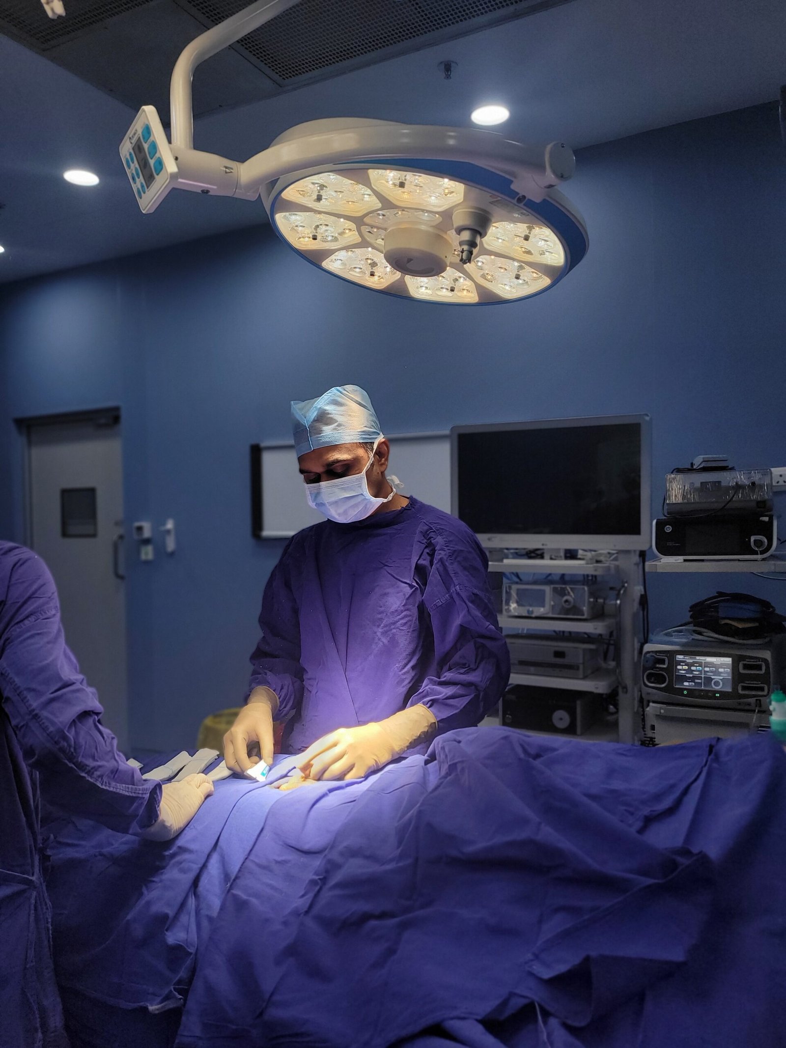
Robotic-assisted partial nephrectomy has become the preferred technique for many high-volume centers around the world. With the da Vinci System, this technique is the least invasive method to complete a partial nephrectomy step. The robotic system is able to provide high quality, three-dimensional images to the surgeon. Additionally, the wristed instruments provide more freedom to the surgeon, as compared to traditional instruments utilized in laparoscopic surgery. Thus, the robotic system is able to provide a surgical level of technical control, allowing the surgeon to excise the tumefaction, perform renal hilar dissection, and perform complex renorrhaphy, as compared to traditional laparoscopy.
Robotic nephrectomy is especially useful when excising complex renal tumefactions. These can include renal tumefactions/ lesions that are located in the renal hilum and are adjacent to the renal vessels or to the renal vasculature, as well as completely endophytic renal tumefactions/ lesiosn that require complex and multi-layered Renal repair after excision. In these specific cases, the systems used in robotic surgery allow the surgeon to complete the repair with great precision and efficiency. This results in improved outcomes in terms of surgical margins, increased renal function post resection, and reduced renal warm ischaemia. Dr. Vikas Singh in Central India performs robotic-assisted partial nephrectomy.
Retroperitoneal vs Transperitoneal Approach
Laparoscopic and robotic partial nephrectomy have two possible access points to the kidney: transperitoneal (via the abdominal cavity) or retroperitoneal (via the retroperitoneal space). The transperitoneal method is better for anterior kidney tumors, upper pole tumors, and cases that require complex reconstruction because it offers better anatomical landmarks and a larger working space. The retroperitoneal method is better for posterior tumors and patients with an abdominal surgical history because of the fewer adhesions. This method is also better for patients that prefer a shorter time from incision to kidney exposure, as there is no abdominal cavity access. As for tumor position, kidney anatomy, and surgical history, Dr. Vikas Singh decides for each specific patient the best method of access.
One of the most patient-beneficial advancements in urological oncology in the past 20 years has been the shift from open to laparoscopic and robotic partial nephrectomy. The benefits impact both the immediate surgical experience and the long-term health outcomes that matter most to patients.
Maximum Kidney Tissue Preservation
The goal of partial nephrectomy is to totally remove the tumour, while preserving as much normal kidney tissue as possible. Laparoscopic and robotic approaches, with their magnified high definition views of the operative field, allow the surgeon to delineate the tumour margin precisely and excise it with the thinnest possible rim of normal tissue, still achieving oncologically adequate negative margins. In certain cases, the improved visualisation in minimally invasive surgery may allow for improved preservation of tissue compared to open surgery, as the magnified view allows for clearer visualisation of the tumour margin compared to the naked eye, thus allowing for a more conservative excision. Every nephron preserved translates directly to improved long-term kidney function.
Minimal Blood Loss & Smaller Incisions
Laparoscopic and robotic partial nephrectomy are associated with significantly less blood loss than open surgery because the pneumoperitoneum (carbon dioxide gas used to inflate the abdominal cavity for working space) exerts mild compression on the operative field, the magnified view allows accurate haemostasis of small vessels before they become major, and the accurate robotic suturing of the kidney reconstruction (renorrhaphy) achieves tight wound closure. Less blood loss is associated with less transfusion, faster post-operative recovery and less blood-related complications. The small port incisions cause dramatically less muscle and tissue trauma, directly reducing post-operative pain and recovery time, compared to the ten to fifteen centimetre flank incision required for open surgery.
Faster Recovery & Shorter Hospital Stay
Patients undergoing laparoscopic or robotic partial nephrectomy are typically discharged two to three days after surgery, compared to four to six days for open partial nephrectomy. Return to light activities may be possible in one to two weeks, return to desk work in two to three weeks, and return to full physical activity in four to six weeks. For open partial nephrectomy, these timelines are generally doubled. For working adults, the self-employed and parents with caring responsibilities, this faster recovery means a much lesser disruption to life during cancer treatment, which is itself an important quality of life benefit alongside the physical advantages.
Better Long-Term Kidney Function Preservation
The improved technical precision of robotic partial nephrectomy , especially in achieving shorter warm ischemia times during kidney clamping , directly leads to better preservation of kidney function in the operated kidney. Warm ischaemia time (the time taken to interrupt the blood supply to the kidney) is one of the most important determinants of post-operative renal function recovery. For every extra minute of warm ischaemia, one per cent of nephron function is lost permanently. Its superior suturing capability enables the robotic platform to perform complex renorrhaphy in less than twenty minutes in the hands of an experienced surgeon, minimising warm ischaemia and maximising functional kidney recovery. In selected cases of exophytic tumours, some surgeons perform a robotic partial nephrectomy (zero-ischaemia) off clamp, eliminating warm ischaemia altogether.
Lower Risk of Chronic Kidney Disease After Surgery
The most significant long-term benefit of partial vs radical nephrectomy , and of technically better partial nephrectomy with maximal preservation of tissue and function , is the markedly reduced lifetime risk of chronic kidney disease (CKD). Published long-term outcome studies have consistently demonstrated that patients undergoing radical nephrectomy have a significantly higher incidence of CKD Stage 3 or worse (eGFR < 60 ml/min/1.73m²), a higher risk of needing dialysis and, critically, a higher overall mortality rate compared to patients undergoing partial nephrectomy, even after adjustment for tumour characteristics. The main driver of this survival advantage of partial nephrectomy is the reduction of cardiovascular mortality of preserved kidney function. CKD is an independent risk factor for cardiovascular disease, myocardial infarction, and stroke, diseases that cause many more deaths among kidney cancer survivors than does the cancer itself in the era of early detection and successful surgical treatment.
Posted onTrustindex verifies that the original source of the review is Google. Dr sahab badiya nature he or samjhate bhi bahut ache se haiPosted onTrustindex verifies that the original source of the review is Google. Dr Vikas Singh Urologist of KDAHOSPITAL is an excellent Doctor. During and after my Operation Dr Singh took personal care. Dr Singh supporting staff are very caring. I recommend patients suffering from UTI, Prostate Gland problems, Kidney Stone, etc to take treatment from Dr Vikas Singh (Retired Senior Professor Pradeep Kundal from Jhabua Madhya Pradesh)Posted onTrustindex verifies that the original source of the review is Google. Bhut achha sir hePosted onTrustindex verifies that the original source of the review is Google. Sir me Mera peostate ka operation kiya tha ab me puri tarah thik hu or mujhe urine bhi bahut ache ata hePosted onTrustindex verifies that the original source of the review is Google. Excellent doctor and great in naturePosted onTrustindex verifies that the original source of the review is Google. Good dr Vikas sirPosted onTrustindex verifies that the original source of the review is Google. 10 mm kidney stone removed via RIRS method, thank you very much Dr Vikas Sir.Posted onTrustindex verifies that the original source of the review is Google. Nice Dr for prostate treatment at kokilaben hospital.
A partial nephrectomy removes the kidney tumour and a small margin of healthy tissue around it , preserving the remainder of the kidney and its function . Radical nephrectomy is removal of the entire kidney. Partial nephrectomy achieves equivalent cancer control, same cure rates, same recurrence rates, same cancer specific survival for T1 tumours (under 7 cm) while preserving significantly better long term kidney function. Partial nephrectomy is the recommended standard of care for T1 tumours when technically feasible, according to international guidelines. Patients who keep their kidney have lower rates of long-term chronic kidney disease, cardiovascular events and overall mortality than those who lose the entire kidney.
Most patients have very good renal function after partial nephrectomy especially when performed with short warm ischaemia time and maximum tissue preservation. The loss of kidney function after partial nephrectomy is dependent on the amount of normal kidney tissue removed (minimised by precise technique), the warm ischaemia time during surgery and the patient’s pre-operative kidney function. In the case of a small peripheral tumour and a short ischaemia time, kidney function is usually only reduced by five to fifteen percent. The remaining kidney compensates over time with mild compensatory hypertrophy and most patients remain in the normal eGFR range in the long term.
Yes, for localised kidney cancer (Stage I and II), partial nephrectomy is curative in the vast majority of patients. Published series show that the 5-year cancer-specific survival for T1a tumours (<4 cm) after partial nephrectomy is >97 to 98 per cent. Partial nephrectomy provides five-year cancer specific survival rates of around 90 to 95 per cent for T1b tumours (4 to 7 cm). The most important determinant of cure is complete excision with negative surgical margins – removal of the whole tumour with a clear rim of normal tissue around it. When appropriate, Dr. Vikas Singh performs intraoperative frozen section margin assessment to confirm complete excision prior to wound closure.
Surgical approach is the key to recovery. Normal hospital stay is two to three days. Light activity can be resumed in one to two weeks. Return to desk work in two to three weeks. Full physical activity in four to six weeks. Hospital stay 4 to 6 days Light activity 2 to 3 weeks Desk work 3 to 4 weeks Full activity 6 to 8 weeks You should not drive for two to three weeks after minimally invasive surgery and four to six weeks after open surgery. A follow-up scan with CT or MRI is planned at 3-6 months to confirm that the tumour has been completely removed and to monitor the kidney.
For most routine partial nephrectomies, both approaches have similar oncologic outcomes. The robotic approach offers certain technical advantages in complicated tumours, central tumours close to the collecting system or renal hilum, endophytic tumours and tumours requiring complex multilayer reconstruction, where the 3D vision, magnification and wristed instrument motion of the robotic system allow for more precise and efficient suturing, shorter warm ischaemia times and better functional recovery. The experienced laparoscopic surgeon can obtain similar results for simple peripheral exophytic tumours. The most important factor for outcomes is always the surgeon’s experience and volume, not the specific technology.
About 10-15% of kidney masses that are managed as suspected cancers on imaging are benign on final pathology, most commonly oncocytoma, angiomyolipoma with minimal fat, or other benign entities. In these cases, the patient does not require further oncologic treatment but can be reassured that no cancer was present, and only needs routine surveillance imaging. In this setting, partial nephrectomy is even more clearly the preferred option, as it would be overtreatment to remove the entire kidney for a lesion that will eventually be found to be benign. Preoperative percutaneous renal mass biopsy is performed in selected cases where imaging is equivocal and a benign diagnosis would change the management plan.
Local recurrence (return of the cancer at the site of the original partial nephrectomy) is infrequent when negative surgical margins are achieved, and in published series, it occurs in about 1-3% of patients at five years for T1 tumours. The rate of distant metastatic recurrence is mainly related to the tumour’s pathological grade, subtype and stage rather than the type of nephrectomy performed (partial vs radical nephrectomy); similar metastatic recurrence rates are observed with both types of nephrectomy. All patients are entered into a structured surveillance programme after partial nephrectomy with regular CT or MRI imaging of the chest, abdomen and pelvis to detect any recurrence at the earliest possible stage when treatment is most effective.
Partial nephrectomy is particularly advocated for tumours less than seven centimetres (T1). Partial nephrectomy for tumours 7–10 cm (T2a) in favourable anatomical settings can be performed by surgeons experienced in this technique in a specialised centre, particularly when there are imperative indications for renal preservation (solitary kidney, bilateral disease, reduced contralateral function). Radical nephrectomy is usually favoured for tumours more than ten centimetres (T2b and T3) because of the very considerable technical difficulties in achieving adequate tumour control and preserving functional parenchyma. Dr. Vikas Singh evaluates each case individually, based on the size and location of the tumour, complexity score and specific kidney preservation requirements of the patient.
No . At present , there is no standard practice for adjuvant chemotherapy , radiation therapy or immunotherapy for completely excised localised kidney cancer ( Stage I and II ) after partial nephrectomy with negative margins . Kidney cancer is resistant to traditional chemotherapy and radiation. There is no evidence at present to support the routine use of adjuvant targeted therapy or immunotherapy after curative surgical resection of localised RCC outside of clinical trials. Dr. Vikas Singh will determine eligibility on a case-by-case basis and coordinate with his medical oncology colleagues for patients who might be appropriate candidates for clinical trial participation Selected patients at high risk (pT3-T4, high Fuhrman grade, sarcomatoid differentiation) may be enrolled in clinical trials of adjuvant therapy.
24/7 Services Available

Copyright © 2026 Urology Center | All Rights Reserved
Design and Developed by Namastetu Technologies Pvt.Ltd