A testicular cancer diagnosis is especially difficult because it happens in young men, typically between 15-35 years old. This population is in the midst of adolescence and developing life goals, relationships, and starting families. Thus, the fear that accompanies a diagnosis of testicular cancer is completely justified.
However, the families of young men afflicted with testicular cancer needs to know this. Testicular cancer is one of the most fully treatable cancers in all of oncology. Even in advanced stages with lymphatic and distant organ metastases, the treatment options and cure rates of testicular cancer are phenomenal. Testicular cancer in stage 1 shows a greater than 99% cure rate. Even in stage 3, the most advanced stage, prognosis of greater than a 70-80% cure rates are astounding in the field of cancer medicine.
The two oval-shaped glands called testes are located in the scrotum and are essential in producing hormones and sperm. Testicular development starts in the abdomen, and the testes usually move down to the scrotum before or soon after birth. Testicular cancer is caused by the same undifferentiated cells found in the scrotum.
Testicular cancer occurs when the germ cells in the testes undergo an uncontrolled mutation and develop a cancerous tumor. Germ cell tumors (GCTs) make up 95% of testicular cancer. Since germ cell tumors can be categorized into sub-classes based on the signs and symptoms, tumor markers, and responsiveness to treatment, their two major classes are called seminomas and non-seminomas.
The strongest association with testicular cancer is a testicle that has failed to descend (cryptorchidism). Both testicles, whether they have had corrective surgery or not, are at a higher risk of developing testicular cancer. Klinefelter syndrome and a family history of testicular cancer also increase the chances.
Any young man with a recent lump or swelling in the scrotum or a change in testicular consistency should have testicular cancer considered in the differential diagnosis – even in the absence of testicular pain. A lack of pain is one of the most misleading and characteristic features of testicular cancer. For this reason, many young men postpone consulting medical practitioners for weeks or even months after the abnormal changes in their bodies. All testicular masses should be assessed without delay by a urologist.
The commonest kind of testicular cancer is seminomas comprised of 55% of germ cell tumors. It arises from germ cells and has a large, uniform population of cells in a tissue sample. It has a good prognosis because it is slow growing and early metastasis is rare. It is very responsive to radiation and cisplatin so treatment is often very effective, even at later stages of metastasis.
These tumors usually present at a later age compared to their non-seminoma counterparts (30 to 45 years). Pure seminomas do not raise alpha-fetoprotein levels which is used a tumor marker for diagnosis and monitoring of treatment. They can slightly elevate human chorionic gonadotropin levels. Seminomas usually spread via the lymphatic system, starting with the retro-peritoneal para-aortic lymph nodes.
Non-seminoma germ cell tumours (NSGCTs) are typically more aggressive than seminomas and spread more easily. They are more prevalent in men aged 15 to 30. Non-seminomas tend to produce more tumour markers than seminomas. The tumour markers include AFP (alpha-fetoprotein) and (sometimes) beta-HCG. These markers assist in diagnosis, aid in monitoring the effectiveness of treatment, and signal any possible tumour recurrence.
Non-seminomas are classified according to the histological components, i.e., the features of the tumour. Most of the pure forms of non-seminomas include embryonal carcinoma, teratoma, yolk sac tumour (endodermal sinus tumour), and choriocarcinoma. Most non-seminomas are mixed tumours and contain choriocarcinoma. Non-seminomas are also highly chemosensitive. The BEP (bleomycin, etoposide, cisplatin) regimen has a cure rate of over 90% in even the most advanced cases of non-seminomas.
Teratoma is a type of non-seminoma germ cell tumour and is classified as a germ cell tumour. It derives from more than one of the three primary embryonic cell layers and so could contain hair, cartilage, and muscle. Teratoma can even contain teeth. Normally, this kind of tumour is classified as mature teratoma and is found to be benign. However, in adults, it can undergo both malignant transformations and differentiation and can even undergo transformation into resistant malignancies, such as carcinoma and sarcoma. Immature teratoma is classified as a malignant tumour. In mature teratoma, certain differentiations are embryonic and primitivised. In the post-chemotherapy retroperitoneal lymph node dissection (PC-RPLND), the residual mature teratoma (which is not responsive to treatment) is surgically excised due to the risk of malignant transformation.
Embryonal carcinoma is the most aggressive type of Pure Non seminomatous type testicular cancer. As is characteristic of all tumours that derive from germ cells, it is composed of primitive embryonic type cells that are comparatively undifferentiated. Embryonal carcinoma has a rapid growth pattern, spreads via lymphatics and blood to distant sites early on, and produces very high levels of serum AFP and, in some cases, beta-HCG. It is the most common element that is found in mixed non-seminomatous tumours. Embryonal carcinoma, despite its aggressiveness is very chemosensitive and therefore, most of the cases are curable even in the metastatic stage with BEP chemotherapy.
In clinic practice, almost all non-seminoma germ cell tumours are not pure subtypes. They are mixed tumours, consisting of different amounts of embryonal carcinoma, teratoma, yolk sac tumour, and, rarely, choriocarcinoma. Managing mixed GCTs involves following non-seminoma protocols. Treatment is mainly guided by the tumour’s clinical stage and the risk classification (good, intermediate, or poor prognosis) of the International Germ Cell Consensus Classification (IGCCC). The IGCCC uses certain tumour markers, clinical stage and histology to place patients in prognostic groups to help determine the level of chemotherapy required.
Testicular cancer is classified using the TNM staging system with an added serum marker component (S stage), which is only used in testicular cancer. This reflects the significance of tumor markers with this cancer. A simplified overview is presented below:
Stage | Cancer Location | Lymph Nodes | Distant Spread | Cure Rate |
Stage 1 | Confined to testis (T1–T4) | None (N0) | None (M0); markers normalise post-orchiectomy | Above 99% |
Stage 2A/B | Any T | Retroperitoneal nodes <2cm (2A) or 2–5cm (2B) | None | 90–95% |
Stage 2C | Any T | Retroperitoneal nodes >5cm | None | 85–90% |
Stage 3A (Good) | Any T | Any N | Lung mets only; markers low-intermediate | 90–95% |
Stage 3B (Intermediate) | Any T | Any N | Lung or non-pulmonary visceral mets; markers elevated | 70–80% |
Stage 3C (Poor) | Any T | Any N | Liver, brain, bone mets; markers very high | 45–55% |
Stage 1 testicular cancer is defined by the cancer being completely localized to the testis and by a complete lack of metastasis to lymphatic tissue, distant organs, and beyond. Following surgical removal of the cancerous testis (orchiectomy), the testicular tumor markers return to the physiological baseline. Stage 1 cancer is divided into subclassifications of 1A and 1B. Classifications of 1A and 1B are differentiated by the absence and presence (respectively) of lymphovascular tumor spread. The optional adjuvant surveillance and/or treatment between Stage 1A and 1B is determined by the classification of the tumor as 1A or 1B.
Both classifications of Stage 1A and 1B testicular cancer seminomas may be treated by active surveillance. 1B testicular seminomas may also be treated by a single-dose regimen of carboplatin and/or para-aortic radiotherapy. Non-seminomatous testicular cancer Stage 1 may be treated by active surveillance, 1 to 2 cycles of combinational chemotherapy consisting of bleomycin, etoposide, and cis-platin (BEP), and/or a surgical procedure to remove the retroperitoneal lymphatic tissue (RPLND). Regardless of treatment type, 1st stage testicular cancer is associated with a >99% long-term cure rate.
Stage 2 testicular cancer has metastasized to the retroperitoneal lymph nodes, the first echelon lymph nodes along the aorta and inferior vena cava in the posterior abdomen. The sub classifications of Stage 2 testicular cancer are largely based on the size of lymph node involvement. When lymph nodes are involved, but are less than 2 cm, the designation is 2A. Nodes between 2 and 5 cm are classified as 2B, while nodes greater than 5 cm are classified as 2C. Stage 2 seminomas are treated with radiation therapy for 2A and 2B, and with three cycles of BEP chemotherapy for 2C and when radiation therapy is not preferred. Stage 2 non-seminomas are treated with three to four cycles of BEP chemotherapy and bilateral RPLND for residual masses.
Stage 3 testicular cancer is when it has metastasized to lymph nodes above the diaphragm or to the lungs (Stage 3A – good prognosis). For Stage 3, BEP chemotherapy will be given systemically, 3 to 4 cycles. Despite Stage 3 testicular cancer involving melanoma, it is still highly curable with a good prognosis involving mainly Stage 3A, with a cure rate of 90 to 95%. This illustrates how germ cell tumors are chemosensitive cancer types and has made testicular cancer a unique case in the field of oncology.
Stage 4 cancer (Stage 3B and 3C in the IGCCC classification) is classified by metastases to distant non-pulmonary organs or body systems such as the liver, brain, or bones, or presence of significantly increased tumor markers. These categories are considered intermediate and poor prognosis. Considering the relatively advanced nature of these presentations, curative outcomes can be expected in most cases. The standard of care is four cycles of BEP chemotherapy. In the case of poor-prognosis patients, high-dose chemotherapy with autologous stem cell rescue may be done at specialized centers. Residual tumors after chemotherapy are reassessed. If they are PCT negative in the case of seminomas, or contain residual teratoma and/or necrosis in the case of non-seminomas, a surgical procedure called post-chemotherapy retroperitoneal lymph node dissection (PC-RPLND) may be required.
The most recognizable sign of testicular cancer is the discovery of a lump, nodule, or swelling in the testicle. Patients make their detection during self-exam or detection can be incidental. Of note is that patients commonly assume that the presence of a lump is associated with pain. However, most testicular masses are associated with testicular cancer and are not painful. Testicular cancer can be dangerous if not addressed in a timely manner.
Every male should self-examine their testicles monthly when the skin is at its most relaxed, such as directly after a hot shower or bath. The testicle should be placed in the palm and rolled in between the fingers and thumb. The testicle should feel solid, smooth, and slightly firm. The epididymis will feel like a solid comma at the back and is separate from the testicle. If there are lumps present, especially if they are hard, and are associated with the testicle, then a medical evaluation should be sought post-haste.
A scrotum that feels heavy, overbearing, or simply feels denser without any apparent painful cause, may be an early sign of testicular cancer. This is due to the increasing weight of the tumor inside the testis. Most will assume this is due to some sort of muscle strain or overexertion, or will brush the symptom off. It is recommended to do a scrotal ultrasound if the heaviness in the scrotum persists for 1 or 2 weeks.
A vague and unclear pain in lower abdomen and groin, especially if it persists and if no association can be made with any physical trauma, can be linked to testicular cancer. This dull pain can be due to the pressure of the tumor, or can also be due to the early involvement of the retroperitoneal lymph nodes with pressing on the surrounding tissues. Also, large retroperitoneal node involvement can be linked to advanced testicular cancer and can cause back pain, which can also be mistaken for a physical back pain. This is especially true in large retroperitoneal node involvement cases.
A hydrocele is the name for the abnormal accumulation of fluid around the testicle within the scrotal sac. It may form due to the reactive response from a testicular tumor. When a hydrocele appears in a young male for the first time, who has no past of trauma or infection, testicular cancer needs to be ruled out by a scrotal ultrasound. This has to be done before any surgical or drainage management of the hydrocele is done. In a few circumstances, a hydrocele could block clinical examination of the testicular tumor. Thus, an ultrasound is vital. For a young male, hydrocele drainage, without having an ultrasound to prove the testicle is normal, is never permissible.
Gynaecomastia means swelling of breast tissue in males and it often occurs as a paraneoplastic syndrome in males with testicular cancer. Swelling of breast tissue occurs as a result of excess synthesis of beta-HCG (human chorionic gonadotropin) by the tumor, especially by the elements of choriocarcinoma. Beta-HCG resembles LH (luteinising hormone) and drives the testes and the adrenal glands to secrete oestrogen, which promotes the augmentation of breast tissue. Any young male who presents with new-onset breast tenderness or breast swelling should undergo testicular examination and scrotal ultrasound as part of his evaluation.
A scrotal ultrasound is an essential first step when diagnosing potential testicular cancer. It is a simple and safe non-invasive procedure that uses sound waves to create images. Nearly all testicular tumors are intratesticular; thus, the ultrasound can test whether there are intratesticular masses, practically defining testicular cancer, almost 100% of the time. The ultrasound can test the size of the intratesticular/extratesticular masses, as well as the blood that is supplied to the masses and the extent and form of the masses, and other information that helps with the surgical plan.
A testicular ultrasound can be completed 24-48 hours after the testicular cancer is suspected, so there should not be delays of weeks. Dr. Vikas Singh at Kokilaben Hospital, Indore, guarantees an ultrasound is done the same day to every patient that is suspected of having testicular tumors.
Three blood tests form the essential tumor marker panel for testicular cancer and need to be taken prior to any surgery (orchiectomy) as both diagnostics and staging require:
these are produced by yolk sac tumor elements in non-seminomas. They are normal in pure seminomas. An elevated AFP in a patient presumed to have pure seminoma means the tumor is actually a mixed GCT (non-seminoma) and thus alters management decisions.
can be mildly elevated in pure seminoma, but markedly so in non-seminomas with trophoblastic elements. Very high beta-HCG suggests choriocarcinoma elements and indicates intermediate/poor prognosis.
this is a non-specific marker of tumor burden. Elevation of LDH stands as a part of the IGCCC risk classification for advanced testicular cancer. A very elevated LDH with other markers suggests a high tumor burden.
When a testicular mass is found on ultrasound and the tumor markers are collected, a CT scan of the abdomen and pelvis – and separately the chest – is performed to stage the disease. The CT scan provides a view of the retroperitoneal lymph nodes (para-aortic and para-caval nodes), the mediastinal and hilar lymph nodes, the lungs, the liver, and other solid organs. All enlarged lymph nodes and/or pulmonary nodules are recorded and measured to document the clinical stage and the post-orchiectomy treatment plan.
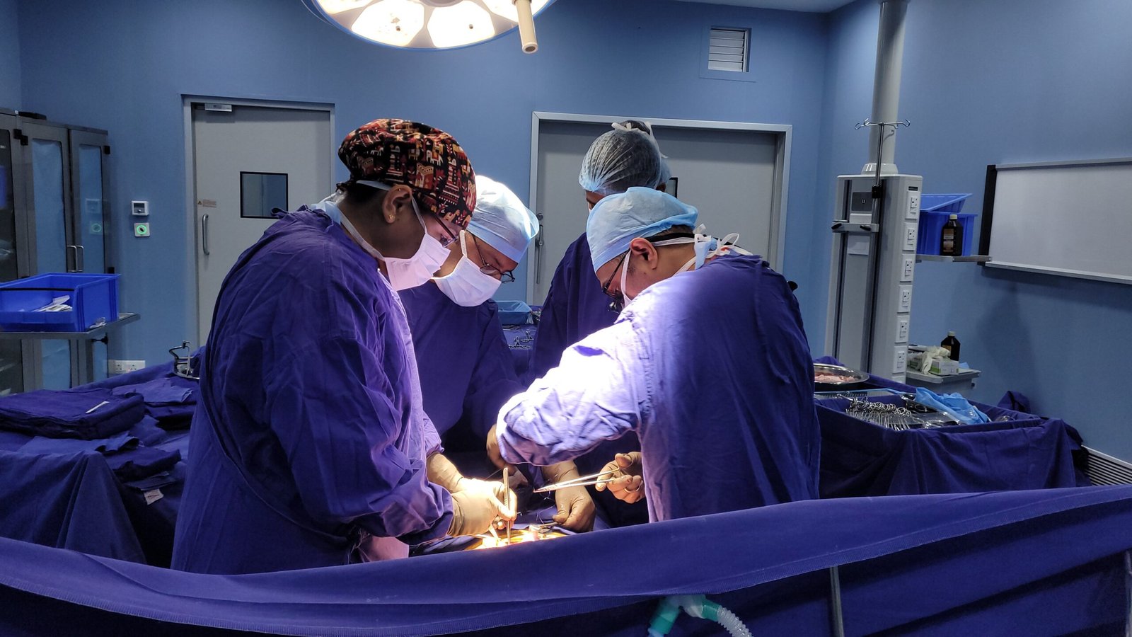
Surgical removal of the entire testicle via the groin (inguinal approach) (Radical orchiectomy) serves a dual purpose in testicular cancer: it is the primary treatment protocol and serves as the definitive diagnostic procedure. Cancer in testicular tissue is suspected only, therefore scrotal access is contraindicated. If the scrotum is opened, the tissue drainage patterns would be altered resulting in potential metastasis of cancer to the testicular lymph tissue. This would complicate both the staging and treatment of the testicular cancer.
General anesthesia is used for the 3 to 5 inch groin incision. The testicle, spermatic cord, and scrotal tissue are removed en masse after the spermatic cord is crushed and ligated within the abdomen at the internal inguinal ring. This technique also has the effect of uterine drainage spread via the abdominal lymph cavities to the retroperitoneal lymph nodes. The procedure generally causes very minimal discomfort and takes 30 to 45 minutes. Most patients return home the next day after the procedure.
FDG-PET/CT scans (fluorodeoxyglucose positron emission tomography) are used for testicular cancer staging as follows:
FDG-PET/CT scans are invaluable in the case of residual masses after chemotherapy for seminomas. For non-seminomas, residual masses can sometimes have non-FDG-avid mature teratoma. In the case of seminomas, residual masses are often necrosis/fibrosis which are PET-negative (do not require any further management) or viable tumors (are FDG-positive and require further management). Thus, FDG-PET/CT scans are crucial for managing post-chemotherapy residual masses for seminomas.
In case of non-seminomas, FDG-PET/CT scans are not as informative as some mature teratomas, which are still required to be excised, can be FDG-negative. For non-seminomas, CT scans remain the most important method of staging.
Understanding the key differences between seminoma and non-seminoma guides every treatment decision after radical orchiectomy. Here is a clear comparison:
Factor | Seminoma | Non-Seminoma |
Age at Presentation | 30–45 years typically | 15–30 years typically |
AFP Level | Never elevated (pure seminoma) | Elevated in majority |
Beta-HCG Level | Can be mildly elevated | Often markedly elevated |
Growth Rate | Slower growing | Faster growing |
Radiation Sensitivity | Highly radiosensitive | Radiation-resistant |
Chemo Sensitivity | Very high (carboplatin, BEP) | Very high (BEP is primary) |
Stage 1 Management | Surveillance, carboplatin, or XRT | Surveillance, BEP x 1-2, or RPLND |
Post-Chemo Residual Mass | PET-CT guides – resect if PET+ | Always resect >1cm mass (PC-RPLND) |
Prognosis (Stage 1) | Above 99% | Above 99% |
When treating patients with Stage 1 testicular cancer (cancer is in the testicle only), following orchiectomy and with normalising tumour markers, it is critical that the treatment burden is minimized. Quality of life must also be considered when planning treatment.
Seminoma Stage 1 Treatment Options:
Stage 1 Non-Seminoma Options:
Systemic chemotherapy becomes the mainstay of treatment for locally advanced testicular cancer, which includes retroperitoneal lymph node involvement or distant lymph nodes (without distant organ metastasis):
Even the most advanced Stage 4 testicular cancer, with metastasis to distant organs, is often curable. Treatment is determined by the IGCCC risk classification:
Fertility preservation is perhaps the most important and overlooked aspect of the management of testicular cancer in young men. Once diagnosed, testicular cancer treatment is usually urgent; however, in the time leading up to radical orchiectomy, it is essential to arrange sperm banking. Delaying this surgery by only 1 to 2 days can allow young men the opportunity to plan for biological children in the future.
Sperm banking is the collection of semen and the cryopreservation of sperm at a fertility laboratory. These sperm can be stored and thawed to be used for IUI, IVF, or ICSI sperm banking for at least 20 years.
There is a clear and important reason why sperm banking is performed before treatment.
After a radical orchiectomy, the vast majority of men maintain their normal testosterone levels as the remaining healthy testicle compensates for the removed testicle. Normally, only one testicle is needed for adequate testosterone production.
However, the body is more than capable of sustaining testosterone levels in a number of other unique circumstances — in men that undergo bilateral orchiectomy (bilateral testicular cancer, for example); in men that had pre-existing low levels of testosterone due to the cancerous testicle; in men that had prolonged, aggressive chemotherapy that causes Leydig cell dysfunction and subsequently low levels of testosterone — levels of testosterone may decrease. Deficiency of testosterone can be characterized by a number of symptoms including but not limited to — increased levels of fatigue, decreased libido, erectile dysfunction, changes in mood, decreased muscle mass, increased body fat, and other symptoms.
Typically, testosterone levels are assessed and measured after 6-12 months of treatment, and then are done in intervals for monitoring. If levels of testosterone are deemed to be low and the symptoms are present, testosterone replacement is done via injections, topical pathways, or transdermal (drugs or hormones absorbed through the skin) patches. Replacement is done without the fear that testicular cancer will reoccur, and improves the quality of life for men that are testosterone deficient. Part of the ongoing and long-term follow up for testicular cancer patients, Dr. Vikas Singh monitors the hormonal levels of his patients.
Posted onTrustindex verifies that the original source of the review is Google. Dr sahab badiya nature he or samjhate bhi bahut ache se haiPosted onTrustindex verifies that the original source of the review is Google. Dr Vikas Singh Urologist of KDAHOSPITAL is an excellent Doctor. During and after my Operation Dr Singh took personal care. Dr Singh supporting staff are very caring. I recommend patients suffering from UTI, Prostate Gland problems, Kidney Stone, etc to take treatment from Dr Vikas Singh (Retired Senior Professor Pradeep Kundal from Jhabua Madhya Pradesh)Posted onTrustindex verifies that the original source of the review is Google. Bhut achha sir hePosted onTrustindex verifies that the original source of the review is Google. Sir me Mera peostate ka operation kiya tha ab me puri tarah thik hu or mujhe urine bhi bahut ache ata hePosted onTrustindex verifies that the original source of the review is Google. Excellent doctor and great in naturePosted onTrustindex verifies that the original source of the review is Google. Good dr Vikas sirPosted onTrustindex verifies that the original source of the review is Google. 10 mm kidney stone removed via RIRS method, thank you very much Dr Vikas Sir.Posted onTrustindex verifies that the original source of the review is Google. Nice Dr for prostate treatment at kokilaben hospital.
Yes, testicular cancer is among the most curable of malignant conditions. The cure rate for stage 1 testicular cancer exceeds 99%. The prognosis for stage 3 (metastatic, “good”) testicular cancer after the administration of cisplatin-based chemotherapy is excellent, and cure rates exceed 90%. Stage 3 (poor prognosis) patients with liver, brain, and bone metastasis have cure rates between 45 and 55%. If you have testicular cancer, the most important thing is: do not wait. Get treatment initiated as soon as possible with a physician who has experience in the management of testicular cancer.
Most men after treatment for testicular cancer maintain their fertility, particularly one who has undergone an orchiectomy for Stage 1 testicular cancer (managed as Stage 1), and has received only one cycle of chemotherapy. Men who need only chemotherapy have one testicle and thus, a testicle frequently produces sperm. Many denoted impotent because of interruption of the chemotherapy cycle regain their ability to produce sperm within two to three years. Patients undergoing a retroperitoneal lymph node dissection (RPLND) are impotent as a result of the surgery, and for this reason, sperm banking is recommended.
Bilateral testicular cancer (having tumors in both testicles at the same time) is an infrequent diagnosis. It comprises only about 2 to 5% of all testicular cancer cases. Generally, when testicular cancer is diagnosed, one testicle is surgically excised. The other, still healthy, testicle will not cease its normal functions. It will continue to produce testosterone and sperm. Very seldom is it possible that a surgically excised testicle is not the only one removed. This can be the result of bilateral synchronous or metachronous tumors, or due to certain genetic syndromes. If that is the case, replacement therapy of testosterone is a necessity with no determined end point.
The surgery is radical orchiectomy, where the surgeon will remove the entire male sex gland through a slit in the groin region (inguinal). The procedure is said to take around 30-45 minutes while the patient is under general anesthesia. The majority of patients can go home the day after. The pain after is said to be moderate at worst and subsides after taking some pain killers. Patients can start moving around lightly a week after surgery. The can start exercising and resuming normal activities after 2-3 weeks. Most patients can resume their normal lives after 3 weeks.
Before administering BEP (Bleomycin, Etoposide, and Cisplatin) regimen, the treating physician should assess the overall clinical picture and health, cost, and benefit/side-effect analyses on the patient, and remain within the policy guidelines for the treatment of the disease. This regimen is associated with multiple side effects: nausea, vomiting, fatigue, alopecia, neuropathy, loss of hearing, reduced fertility, increased vulnerability to infections due to leukopenia. Specific to bleomycin, there is some form of pulmonary toxicity, due to bleomycin-induced lung damage, and so lung function tests are monitored in BEP. Most side effects are temporary; hearing loss and some neuropathies can be long lasting.
Yes – relapse rates depend on the disease stage and the treatment delivered after each stage, and they each have a range of expected relapse rates. After Stage 1 and monitored with Active Surveillance, 15 to 30% relapse. Nearly all of this cohort of Stage 1 relapsed patients are further cured with chemotherapy. After Stage 1 and treatment with Adjuvant Chemotherapy, only 3 – 5% of that cohort goes on to relapse. After chemotherapy for Stage 2-3 disease, 20 to 30% of that cohort relapses. Following chemotherapy administered for the relapsed disease, there are good success rates for Salvage Chemotherapy. The best way to detect a relapse early is to implement regular follow-ups with the indicated CT scans and tumor marker tests. You must keep up with your surveillance.
Yes – we can integrate a testicular prosthesis (a cosmetic replacement for a removed testicle) during an orchiectomy or as a distinct procedure in the future. The prosthesis is a saline silicone implant which reconstructs the appearance of a normal scrotum on the outside and the inside. The prosthesis contributes nothing to the body in terms of hormone production or sperm. It is a cosmetic device meant to make the scrotum appear whole. Many men seek a prosthesis for psychological or body image reasons. Dr. Vikas Singh reviews this option with every patient prior to orchiectomy and if the patient consented, places the prosthesis at the same time.
Yes, almost all men have normal sexual function after radical orchiectomy. The body has a remarkable ability to adjust to the presence of only one testicle, and the testosterone from that one testicle is usually enough to maintain sexual desire and the ability to have erections and ejaculatory orgasm. There is one notable exception to this, and that is RPLND (Retroperitoneal lymph node dissection). If the surgery is not performed using a technique that is “nerve-sparing,” it may cause what is known as “retrograde ejaculation.” In the current state of the art, about 90 to 95 percent of patients will retain normal ejaculation in the antegrade direction after surgery. Dr. Vikas Singh has thorough discussions regarding the anticipated sexual function that may be expected with all of the treatment options prior to any actual procedure.
Testicular self-exams are suggested for males starting around age 13 to 14, which for most begins with puberty, and should be done monthly. After a warm shower, with the relaxed skin of the scrotum, cup the testicle with the palm and roll it, and check for hard lumps, size changes, or changes in consistency. The structure running along the back of the testicle is the epididymis, and is not a tumor. Any hard lumps or changes in the testicle should be evaluated by a doctor.
24/7 Services Available

Copyright © 2026 Urology Center | All Rights Reserved
Design and Developed by Namastetu Technologies Pvt.Ltd