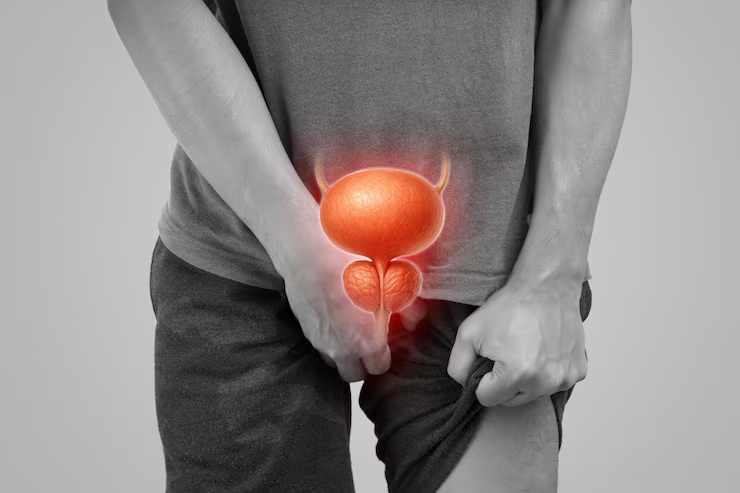
Most people know about the kidneys and the bladder. Fewer still have heard of the ureter, the slender muscle tube that joins the two. But some of the most acutely painful and potentially dangerous urological conditions are centred around the ureter. Some of the worst pain the human body can experience is caused by a stone lodged in the ureter. If untreated, an obstructed ureter will slowly destroy the kidney above it. Every urinary infection will damage the developing kidney of an undiagnosed child with a refluxing ureter.
The ureter is the bottleneck of the urinary tract and is very small, barely four to seven millimetres in diameter at its narrowest points . It is at these naturally narrow points that stones lodge, where strictures form, and where developmental abnormalities cause obstruction. These are conditions requiring specialist urological assessment and in many cases surgical intervention to preserve renal function and relieve pain.
The ureters are two narrow, muscular tubes, one on each side of the body, that carry urine from the kidneys to the urinary bladder. In the adult, each ureter is 25–30 cm long, passing from the renal pelvis (the funnel-shaped collecting area in the centre of the kidney) inferiorly along the spine in the retroperitoneal space, crossing the pelvic brim and entering the posterior wall of the bladder at the trigone.
The ureter is not a simple drainage pipe. It actively moves urine to the bladder in a process called peristalsis. The smooth muscle of the ureteric wall contracts in coordinated waves about one to five times per minute, propelling small boluses of urine along the tube in any position of the patient’s body. This muscular activity explains why ureteric colic from a stone is so severe, the smooth muscle violently contracts against the obstruction and produces intense waves of pain following the ureteric path from the flank to groin.
The ureter has three normal sites of narrowing where stones, blood clots and other material most commonly become impacted. These are the pelviureteric junction (PUJ) where the renal pelvis joins the ureter, the crossing of the pelvic brim and the vesicoureteric junction (VUJ) where the ureter enters the wall of the bladder. The VUJ is the narrowest part and the most common site for stone impaction. At the VUJ the ureter passes through the wall of the bladder muscle at an oblique angle, this oblique tunnel forms a one way valve mechanism (the vesicoureteric valve) which normally prevents urine refluxing backwards from the bladder into the ureter during bladder contraction. Vesicoureteral reflux (VUR) occurs when this valve mechanism does not work.
The ureter is the only conduit between kidney and bladder on each side; therefore any significant obstruction to the ureter will have immediate and profound effects on the kidney above. Urine that can’t drain collects in the renal pelvis. This causes progressive dilation and distension of the renal pelvis (hydronephrosis) . This raises pressure within the kidney and if left for a period of time will progressively destroy nephron function. The kidney is surprisingly tolerant of short term obstruction, but is susceptible to progressive irreversible injury with long standing obstruction. Prompt recognition and relief of ureteric obstruction is therefore vital to protect the kidney.
Ureteric Stone (Stone Stuck in the Ureter)
Stone that has migrated from the kidney into the ureter causing sudden, severe colicky flank pain radiating to the groin, nausea and haematuria. Stones <5 mm often pass spontaneously with fluids and pain relief. Larger stones or stones that cause infection, obstruction or ongoing pain need ureteroscopic laser removal, a keyhole endoscopic procedure done through the urethra.
VUR is abnormal retrograde flow of urine from the bladder into the ureter and up to the kidney during bladder contraction. It is most common in children and causes febrile urinary tract infections that damage developing kidney tissue with each episode. VUR is classified from I-V according to severity and may resolve spontaneously with age or require endoscopic or surgical correction.
PUJO is a narrowing or obstruction at the junction between the collecting system of the kidney and the ureter that prevents urine from draining freely, leading to progressive hydronephrosis (swelling of the kidney). It is the most frequent cause of important hydronephrosis in children . Laparoscopic pyeloplasty is the surgical reconstruction of PUJ and provides definitive and very successful treatment.
Ureteric conditions present with distinct and recognisable symptoms, many of which warrant immediate medical intervention. Please do not ignore the warning signs below.
Severe Flank Pain Radiating to Groin (Ureteric Colic)
Sudden Severe Flank or Loin Pain Not Settling With Painkillers
Same day emergency urological assessment is required in acute ureteric colic not adequately controlled by standard oral analgesics or preventing normal function. Uncontrolled pain per se is an indication for hospital-based management including intravenous analgesia, imaging to confirm stone position and size, assessment for concurrent infection and planning for intervention if spontaneous passage is unlikely. If the pain is severe, do not allow it to settle by itself, complications such as infected obstruction can develop quickly.
Any episode of visible blood in the urine , whether painful or painless , requires urological evaluation . Haematuria together with ureteric colic and pain is highly suggestive of a stone . Painless haematuria may be due to a ureteric, bladder or kidney tumour and should never be dismissed as minor without proper investigation including CT urogram and cystoscopy. Referral is warranted after a single episode.
Fever >38 degrees Celsius with flank pain is a urologic emergency until proven otherwise. The combination is suggestive of an infected obstructed upper urinary tract which can progress to life threatening sepsis within hours. Emergency assessment includes blood cultures, intravenous antibiotics and urgent decompression of the obstructed system , either by retrograde ureteric stent insertion (DJ stent) or percutaneous nephrostomy (drainage tube placed directly into the kidney through the skin). If you have this combination of symptoms, you should go straight to the emergency department.
In any imaging study in which a dilated ureter (hydroureter) or dilated kidney collecting system (hydronephrosis) is seen , even in an asymptomatic patient , assessment by a urological specialist is mandatory. Asymptomatic hydronephrosis may be a silent obstruction which is damaging the kidney progressively. The cause, whether stone, stricture, tumour, PUJO or external compression has to be identified and appropriate management initiated. The finding of a dilated ureter on imaging should never just be ‘watched’ without further assessment of the underlying etiology.
Vesicoureteral reflux should be investigated in any child who has had two or more febrile urinary tract infections, with fever, loin pain, and a positive urine culture . VUR allows bacteria-containing urine from the bladder to reflux into the kidney during urination, resulting in recurrent kidney infections (pyelonephritis) that slowly scar the renal parenchyma. Hypertension, reduced renal function and chronic renal disease in adult life result from renal scarring acquired in childhood. Early recognition and treatment of significant VUR by VCUG (voiding cystourethrogram) prevents this accumulation of kidney damage.
Fetal hydronephrosis, the dilation of the kidney’s collecting system seen on the routine fetal anomaly ultrasound scan performed during pregnancy, is one of the most common antenatal findings, occurring in about 1–2% of all pregnancies. A planned schedule for post-natal assessment should be arranged with a paediatric urologist or paediatric nephrologist before birth. Post-natal ultrasound, VCUG (if VUR suspected) and MAG3 nuclear medicine scan post delivery to assess degree of obstruction and differential renal function. The majority of cases of mild antenatal hydronephrosis resolve spontaneously but significant PUJO or VUR detected on post-natal investigation requires timely intervention. Dr Vikas Singh views antenatal referrals from the time of diagnosis of anomaly scan to plan postnatal assessment.
The DJ stent, or double-J stent, is one of the most commonly used devices in urology. It is one of the most common urological devices inserted, yet many patients that have one inserted have little understanding of what it is, why it is there, and more importantly, why it needs to be removed in a timely fashion. This section includes a full explanation in patient-friendly language.
Aspect | Details |
What it is | A soft, flexible hollow tube with a curled (pigtail) end at each extremity , one curl in the kidney, one in the bladder |
Material | Medical-grade polyurethane or silicone , body-compatible, smooth surface to reduce irritation |
How it works | Bypasses any obstruction in the ureter, keeping urine flowing from kidney to bladder even when the natural passage is blocked |
Inserted how | Cystoscopically , passed through the urethra and bladder up into the ureter and kidney under local or general anaesthesia. No external incisions. |
When it is needed | After ureteroscopic stone removal; treating ureteric obstruction; after ureteric surgery or injury; temporary drainage before definitive surgery |
Duration in body | Typically 2–6 weeks; up to 3 months maximum; must not be left longer without planned removal or exchange |
Common side effects | Urinary frequency and urgency; mild haematuria; flank discomfort when voiding; occasional bladder discomfort |
Removal | Cystoscopic removal , a short outpatient procedure. Some stents have a string visible at the urethral meatus for self-removal or GP removal |
Forgotten stent risk | An overstayed stent encrusts with mineral deposits and may require complex removal. Always maintain follow-up for stent removal. |
Ureteric conditions in children differ importantly from adult disease both in the conditions that occur most frequently and in how they are managed. Ureteric conditions in children are most often congenital, being present from birth as the result of abnormal development of the urinary tract during foetal life, rather than acquired as in adults. Early recognition and management of congenital ureteric abnormalities is important, because in infancy and early childhood the kidneys are still developing and obstruction or reflux left untreated at this age leads to permanent kidney damage, which is carried throughout the child’s life.
Condition | Age of Detection | Typical Presentation | Preferred Treatment |
Antenatal hydronephrosis | Before birth on anomaly scan | Dilated renal pelvis on Fetal ultrasound | Monitor post-birth; VCUG if VUR suspected; pyeloplasty if PUJO confirmed |
PUJO | Newborn to adolescence | Antenatal detection or recurrent loin pain/UTI | Laparoscopic pyeloplasty , gold standard; minimal recovery |
VUR | Any age; often < 5 years | Febrile UTI; recurrent UTI in child | Medical: prophylactic antibiotics. Surgical: endoscopic STING/HIT injection or ureteric reimplantation |
Ureteric stone | School age and above | Sudden severe pain; haematuria; vomiting | Most pass spontaneously; ureteroscopic laser removal if needed |
Duplex collecting system | Antenatal or post-natal | Recurrent UTI; obstruction of upper pole moiety | Conservative surveillance; endoscopic or surgical management of complications |
Dr Vikas Singh offers complete paediatric urological treatment for ureteric conditions starting from antenatal counselling consultation when hydronephrosis is detected on the anomaly scan, to postnatal investigation and surveillance, laparoscopic pyeloplasty for PUJO, endoscopic VUR treatment and ureteroscopic stone management in older children. We welcome parents of children with known or suspected ureteric conditions for consultation and will offer clear and honest advice as to the diagnosis, natural history of the condition and the treatment options available.
Laparoscopic pyeloplasty for PUJO in children , the preferred treatment for pelvi-ureteric junction obstruction , is performed under general anaesthesia through small incisions of less than one centimetre, with children usually discharged within two to three days and resuming normal activity within one to two weeks. Success rates are >95% with experienced hands and the vast majority of operated kidneys have improved drainage and preserved or improved renal function on follow-up nuclear medicine scanning.
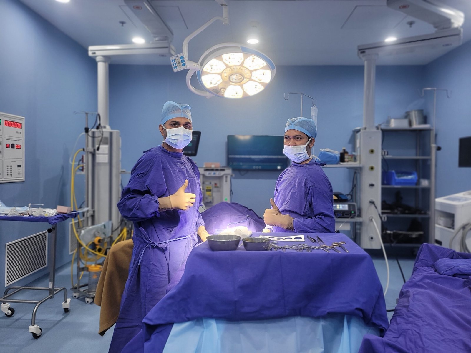
Ureteric disease covers the whole spectrum of urological urgency, from the acute emergency of an infected obstructed ureter to the long-term surveillance of a child with mild VUR. Surgical skill and clinical judgment are needed to know when to intervene urgently, when to wait and which procedure is best for each patient. Here’s why patients from all over Indore and Central India choose Dr. Vikas Singh:
Posted on GoogleTrustindex verifies that the original source of the review is Google. अत्यंत ज्ञानवर्धक ज्ञान मिला धन्यवाद, क्या Dilo dil D के सेवन से नाक बंद होने की समस्या होती हैPosted on GoogleTrustindex verifies that the original source of the review is Google. Dr vikas singh sir ne 24 mm ka stone kokilaben ambani hospital me high frequency wali RIRS method se bahut ache se bina Dard ke nikal diya or aaj DJ Stent bhi removal kr diyaPosted on GoogleTrustindex verifies that the original source of the review is Google. We are incredibly grateful to Dr. Vikas. My mother underwent a URS surgery for a stone under his care, and he did a fantastic job. Not only is he an expert in his field, but he is also extremely humble and reassuring. My mother is completely healthy and comfortable now. Thank you, Dr. Vikas, for your wonderful treatment and care.Posted on GoogleTrustindex verifies that the original source of the review is Google. Meri ka 20mm or 15 mm ka stone tha or hum log Dhar se hain or dr vikas singh sir ne kokilaben hospital me kidney stone mini PCNL method se stone remove kr diya hai thanks dr vikas sirPosted on GoogleTrustindex verifies that the original source of the review is Google. Meri mother ka name fulwanti he, hum indore se hai dr vikas singh sir ne kokilaben hopital me 21 mm ka stone nikal diya ab meri mother bilkul thik haiPosted on GoogleTrustindex verifies that the original source of the review is Google. Dr. Vikas Singh was very prompt with his treatment for my father. The entire consultation was extremely helpful, and he explained everything clearly. We are very satisfied with the care and guidance provided by him.Posted on GoogleTrustindex verifies that the original source of the review is Google. अभी आपका AToZ नही देखा है लेकिन उपाय वाला उपयोगी है साधुवाद।Posted on GoogleTrustindex verifies that the original source of the review is Google. 25 mm stone removed through mini PCNL therapy safely … I’m from sagar Mp and experience was good… nice doctor and associated staffPosted on GoogleTrustindex verifies that the original source of the review is Google. मैंने अपनी दोनों किडनी की पथरी का ऑपरेशन RIRS (Retrograde Intrarenal Surgery) विधि से डॉ. विकास सिंह सर के मार्गदर्शन में करवाया। ऑपरेशन से पहले मैं और मेरा परिवार काफी चिंतित और डरे हुए थे, लेकिन जब हम डॉ. सर से मिले तो उनकी स्पष्ट सलाह, आत्मविश्वास और सकारात्मक व्यवहार ने हमारा सारा डर दूर कर दिया। सबसे बड़ी बात यह रही कि मुझे केवल एक दिन के लिए अस्पताल में भर्ती रहना पड़ा और अगले ही दिन डिस्चार्ज कर दिया गया। ऑपरेशन के तुरंत बाद पथरी के दर्द से काफी राहत मिल गई, जो हमारे लिए किसी चमत्कार से कम नहीं था। डॉ. विकास सिंह सर का व्यवहार अत्यंत विनम्र, सहयोगपूर्ण और भरोसेमंद है। साथ ही अस्पताल का पूरा स्टाफ भी बहुत पेशेवर, संवेदनशील और मददगार है। कोकिलाबेन अस्पताल में हमें उत्कृष्ट चिकित्सा सुविधाएँ और बेहतरीन सेवा प्राप्त हुई। शुरुआत में हमें यह चिंता थी कि कहीं हम गलत जगह तो नहीं आ गए हैं, लेकिन आज अपने सफल उपचार के बाद मैं पूरे विश्वास के साथ कह सकता हूँ कि यह हमारा बिल्कुल सही निर्णय था। यदि आप किडनी स्टोन की समस्या से परेशान हैं और कम समय में सुरक्षित एवं प्रभावी उपचार चाहते हैं, तो मैं निःसंकोच डॉ. विकास सिंह सर की सलाह दूँगा। उनके अनुभव, विशेषज्ञता और मरीजों के प्रति समर्पण पर आप पूर्ण विश्वास कर सकते हैं। धन्यवाद, डॉ. सर और पूरी टीम, जिन्होंने मुझे दर्द से राहत देकर एक नई शुरुआत दी।Posted on GoogleTrustindex verifies that the original source of the review is Google. Mere father ka prostate ka ilaj dr vikas sir ne kiya or sir ka nature bahur acha hai
The ureter is the narrow muscular tube (about 25–30 cm long, 4–7 mm wide) that connects each kidney to the bladder , one on each side. It pushes urine actively from the kidney to the bladder using peristaltic muscular contractions. The ureter is critically important because it is the only drainage route for the kidney, and any significant obstruction of the ureter causes urine to back up into the kidney (hydronephrosis), raising pressure and progressively destroying kidney function if the obstruction is not relieved. There are three sites of natural narrowing within the ureter where stones most commonly get stuck and where developmental narrowing (such as PUJO) most commonly occurs.
Ureteric colic caused by a stone has a characteristic pain pattern that helps differentiate it from the majority of other causes of abdominal or back pain. The pain has a sudden onset (no warning), is severe (colicky) in nature, occurs in waves and there is no comfortable position. The pain is felt in the flank and radiates towards the groin, lower abdomen, inner thigh or genitalia. There is usually associated nausea, vomiting and visible blood in the urine. There is no fever or rigors unless the stone is also causing infection. If you have this symptom pattern you should seek urgent medical assessment , a plain X-ray or CT scan can confirm the diagnosis within minutes .
Yes , many ureteric stones spontaneously pass without surgical intervention. Stone size is the main factor influencing the probability of spontaneous passage. Spontaneous passage occurs in approximately 80 percent of stones less than 4 mm in size. About 50 to 60 percent of stones between 4 and 6 mm pass. Stones >6 to 7 mm infrequently pass spontaneously and usually require surgical intervention. The location of the stone matters too; stones in the upper ureter have a harder time passing than those near the bladder (distal ureter). Often alpha-blocker drugs (tamsulosin) are prescribed to relax the smooth muscle in the ureter and help passage. Good hydration and pain control while awaiting passage is important. Stones that cause fever, uncontrolled pain that does not respond to medication, complete obstruction or obstruction of a solitary kidney do not wait for spontaneous passage and require prompt intervention.
A DJ stent is a soft, flexible hollow tube with a curled end at each end, one curl in the kidney and one in the bladder, which bypasses any obstruction in the ureter and keeps urine draining from kidney to bladder. It is introduced by cystoscopy through the urethra, either under local or general anaesthesia. DJ stents are temporary devices and must be removed or exchanged within their intended dwell time, typically two to six weeks (occasionally up to three months for certain indications). A stent left in place longer than its allotted time can become coated with mineral deposits and may require complicated procedures to remove. Before your doctor places a stent, always have a confirmed stent removal appointment.
Vesicoureteral reflux (VUR) is the retrograde flow of urine from the bladder into the ureter and kidney during bladder contraction due to a defective vesicoureteric valve mechanism. It is graded I to V. Grade I–II (mild reflux which does not reach the kidney) often resolves spontaneously with growth of the child. Grade III (reflux to kidney without dilation) may resolve over time with antibiotic prophylaxis. These grades are less likely to resolve and are more likely to cause kidney damage. Grade IV-V (severe reflux with significant kidney dilation) more often need endoscopic (STING injection) or open surgical correction. The management decision depends on the grade of VUR, the age of the child, whether there are break through UTIs despite antibiotics and whether any scarring or functional impairment of the kidney is developing. Individual assessment determines the most appropriate management and not all children with VUR require surgery.
Pelvi-ureteric junction obstruction (PUJO) is a narrowing or blockage of the junction of the collecting system of the kidney (renal pelvis) and the ureter which prevents adequate drainage of urine and causes progressive hydronephrosis (swelling of the kidney). It is the most common cause of significant hydronephrosis in children but can also occur in adults. The definitive treatment is pyeloplasty, which is the surgical reconstruction of the narrowed PUJ into a wide unobstructed junction. Laparoscopic pyeloplasty is the gold standard approach in both children and adults, performed through small keyhole incisions with success rates over 95 percent and a much quicker recovery than open surgery. Operating Time: 2-3 hoursHospital Stay: 2-3 daysReturn to Normal Activities: 1-2 weeks
Yes , PUJO and VUR can cause permanent kidney damage if not treated properly or at all . In PUJO the collecting system is chronically dilated (hydronephrosis). The elevated pressure within this distended system progressively destroys nephron function over months to years and results in renal damage. The damage is particularly rapid and severe in the neonates and infants whose kidneys are still developing. VUR leads to recurrent urinary tract infections . With each infection , bacteria reflux from the bladder into the kidney , leading to pyelonephritis ( kidney infection ) that scars the renal parenchyma . The consequence of renal scarring acquired in childhood may be hypertension, proteinuria and chronic kidney disease in adulthood. Both conditions are very treatable if diagnosed early, which is why it is so important to promptly investigate antenatal hydronephrosis, febrile UTIs in children and incidental hydronephrosis on imaging.
Yes, laparoscopic pyeloplasty is safe and well established in children and even in infants as young as a few months of age in experienced pediatric urologic centers. The procedure is performed under general anaesthesia by a paediatric anaesthesiologist using instruments specially sized for small patients. The keyhole incisions (usually three ports of 3–5mm in infants and young children) cause minimal trauma and children recover very quickly. Most children are tolerating oral feeds within hours of surgery and ready for discharge within two to three days. Success rates are comparable or better than open pyeloplasty with the advantage of much smaller scars and quicker recovery.
Pyonephrosis (infected obstructed kidney): This is a urological emergency. If a stone is in the ureter, it can cause infection as bacteria get trapped in the obstructed urine above the stone. This is a life-threatening condition that requires immediate hospitalisation, intravenous antibiotics and urgent drainage of the infected obstructed kidney by cystoscopic retrograde stent insertion (placing a DJ stent past the stone) or percutaneous nephrostomy (inserting a drainage tube directly into the kidney through the skin under ultrasound guidance). It is generally not appropriate to attempt definitive stone removal at the same time as the infection is being treated, the priority being drainage, antibiotics and stabilisation. Once the infection has settled (usually after two to four weeks) the stone is dealt with definitively.
24/7 Services Available

Copyright © 2026 Dr Vikas Urologist | All Rights Reserved
Design and Developed by Namastetu Technologies Pvt.Ltd