
A kidney stone can strike without warning, a wave of pain that starts in the back and radiates toward the groin and can bring some patients to their knees. Sometimes a kidney stone is found completely by chance on an ultrasound scan with no symptoms at all. The approach to treatment whether silent or agonising should always be the same: proper evaluation, appropriate treatment for the stone and critical investigation into why the stone formed so it does not come back.
Kidney stones are one of the commonest urological conditions . About 10 to 15 percent of the people will have them during their lifetime . They show a strong tendency for recurrence . The high prevalence in the Central India region is largely due to the hot climate, relatively low fluid intake and dietary patterns. The good news is that over 90% of kidney stones can be treated in a complete and safe manner, most without open surgery, with modern techniques such as laser lithotripsy and PCNL that allow same-day or next-day discharge.
A kidney stone is a hard, crystalline deposit of minerals and salts that forms inside the kidney when urine becomes supersaturated, that is, it contains more dissolved minerals than can remain in solution. If the concentration of some minerals in the urine becomes greater than their solubility, then crystals may start to form and aggregate and finally produce a stone that can be as tiny as a grain of sand or as large as a golf ball. The stone is in the kidney to begin with , it may remain there silently for months or years , or it may move down the ureter resulting in the excruciating pain of renal colic .
How Kidney Stones Form Inside the Kidney
Stones form when urine becomes supersaturated with mineral salts, most commonly calcium oxalate. Crystals start to nucleate in the collecting ducts and crystal aggregation occurs over weeks to months. This is helped by a lower volume of urine, changed pH of the urine or lower levels of endogenous inhibitors (citrate, magnesium).
Kidney stones can be anything from sub-millimetre gravel to staghorn calculi filling the entire collecting system . Size is a direct indicator of management, stones less than 5 mm often pass spontaneously, while those greater than 10 mm almost always require intervention. “If left untreated, staghorn stones can grow over time and eventually destroy kidney function altogether.
Yes many kidney stones can be silent in the kidney for months or years. But a ‘silent’ stone is not necessarily a safe one. It can grow, migrate and block the ureter, cause recurrent infection or progressive damage to the kidney by back pressure. All recognised renal calculi should be assessed and followed regardless of symptom status.
Almost always the formation of kidney stones occurs from a combination of factors . Diet, metabolism, environment and genetics all work to produce a urine that is supersaturated with stone forming minerals . Effective prevention is based on knowing the causes for each individual patient.
Low Water Intake & Chronic Dehydration
Inadequate fluid intake is the single most important modifiable risk factor for kidney stones. Low fluid intake produces concentrated urine with high mineral content, dramatically increasing stone formation risk. In hot climates like Central India, sweat losses further concentrate the urine. Increasing fluid intake to produce at least two litres of urine daily reduces stone recurrence by approximately 50 percent , the most evidence-based, cost-effective intervention in stone prevention.
Diet composition has a significant effect on urine chemistry. High oxalate foods (spinach, nuts, chocolate, tea, beetroot) increase urinary oxalate and predispose to calcium oxalate stone formation. Excess sodium increases urinary calcium excretion and decreases citrate, a natural stone inhibitor. High animal protein increases urinary uric acid, calcium and oxalate and decreases citrate and urinary pH. High purine diets (red meat, organ meats, shellfish) increase uric acid levels, which lead to uric acid stones. The cornerstone of prevention of dietary stones is a balanced diet high in vegetables and fluid, moderate in animal protein and low in salt.
Struvite stones (magnesium ammonium phosphate) are the only kind of stone which arises from urinary tract infections caused by urease-producing bacteria (Proteus, Klebsiella, Pseudomonas). These bacteria hydrolyse urinary urea to ammonia, alkalinising the urine and creating ideal conditions for struvite precipitation. Struvite stones grow quickly and can fill the entire renal collecting system (staghorn calculi) and recur if the underlying infection is not eradicated permanently. Long term management necessitates complete stone removal and antibiotic therapy guided by the causative organism.
Primary hyperparathyroidism Excess activity of the parathyroid glands causing increased production of PTH . Increased serum calcium Increased urinary calcium excretion (hypercalciuria) leading directly to calcium stone formation. It occurs in approximately five percent of patients with recurrent stones. Serum calcium and PTH should be evaluated in all patients with recurrent calcium stones. Other metabolic causes are renal tubular acidosis (which produces alkaline urine and low citrate), hyperoxaluria (primary genetic forms or secondary to bowel disease), hyperuricosuria and cystinuria. Besides general measures to prevent stones, these conditions need specific medical management.
A positive family history of kidney stones roughly doubles the lifetime risk for an individual. Cystinuria (cystine stones) Primary hyperoxaluria (rare but severe, calcium oxalate stones from early childhood) Dent’s disease Familial hypercalciuria Genetic conditions directly causing stone disease include: The tendency to form stones is familial even in the absence of these specific inherited conditions, reflecting shared dietary habits, shared environmental exposures, and shared polygenic predisposition to urinary chemistries that favour stone formation. Metabolic evaluation of stone type should be done in men with a first-degree relative with kidney stones even after a first stone episode.
Severe Flank Pain Radiating to Groin (Renal Colic)
Renal colic is one of the worst pains the human body can experience; many patients describe it as worse than childbirth or major surgery. It begins as a sudden, severe, cramping pain in the flank (back, below the ribs on one side), usually radiating around the abdomen to the groin, inner thigh and genitalia, along the ureter’s path. The pain is in waves , it is intense for a few minutes , then it eases slightly , then returns . This is due to the peristaltic contractions of the ureter against the stone causing the obstruction . Unlike pain from a muscular injury (which is worsened with movement), renal colic is characterised by the inability of the patient to remain still, they pace, writhe and cannot find any comfortable position. Such an episode should be urgently assessed as an emergency.
Blood in the urine ( haematuria ) occurs in about 85% of attacks of kidney stones . It is caused by the abrasive effect of the stone on the mucosa of the ureteral or renal pelvis during its migration. The blood can turn the urine red or pink when viewed with the naked eye, or dark brown or cola-coloured if there is a large amount of blood and it has been in the urinary tract for a while. Microscopic haematuria, blood detectable only on urine dipstick testing, is even more common. Haematuria in the context of flank pain is highly suggestive of a stone but any haematuria without pain should be investigated to exclude other causes including bladder cancer.
Nausea and vomiting are very common in severe renal colic and occur in the majority of patients with significant ureteric obstruction. There are common neural pathways between the ureters and the gastrointestinal tract through the coeliac and mesenteric plexuses. Intense ureteric peristalsis against an obstruction triggers a visceral reflex that activates the vomiting centre. It is a neurogenic response to the intensity of ureteric spasm. Vomiting with an episode of pain is not a sign of a gastrointestinal problem. It also helps to dehydrate patients who are already producing concentrated stone-forming urine.
Fever with renal colic is a urological emergency If bacteria are present in urine that cannot drain freely due to an obstructing stone, a condition called pyonephrosis (infected obstructed kidney), the infection rapidly ascends and can progress to septicaemia and septic shock within hours. This presentation, flank pain, fever > 38°C and rigours (shaking chills) requires immediate emergency hospital admission, intravenous antibiotics and urgent decompression of the obstructed kidney by either retrograde ureteric stenting or percutaneous nephrostomy. In the setting of acute infection stone removal is dangerous and should not be performed, drain first, treat the stone later when the infection is cleared.
As the stone passes into the lower ureter and the vesicoureteric junction (where the ureter joins the bladder) it may simulate a urinary tract infection with burning or stinging on urination, increased frequency and urgency of urination, and suprapubic discomfort. These symptoms are due to the stone at the VUJ stimulating the trigone of the bladder which is innervated by the urethra . A urine dipstick test and a CT KUB (non-contrast CT of the kidneys, ureters and bladder) should be performed to differentiate the two conditions, as antibiotic treatment alone will not remove the stone, many patients and doctors misdiagnose this as a UTI.
A stone in the vesicoureteric junction, at the bottom of the bladder where it enters, causes a constant feeling of urgency to urinate, even when the bladder is not very full. This is because the affected stone directly stimulates the stretch receptors of the trigone which normally signal bladder fullness. When going to the toilet only a small amount of urine is passed even though a strong urge is felt. This pattern of frequent urgency but low output is a characteristic and clinically useful sign of a distal ureteric stone and should lead to imaging assessment rather than simple antibiotic treatment.
A large proportion of kidney stones are discovered incidentally, on an ultrasound, ct scan or x-ray performed for an unrelated reason, in a patient who has no pain, no haematuria and no urinary symptoms. These ‘silent’ stones could have been there for months or years without any problem. BUT FINDING A SILENT KIDNEY STONE IS NOT A REASON TO BE REASSURED AND DO NOTHING , IT IS A REASON TO BE EVALUATED . The stone needs to be characterised (size, position, composition if possible), kidney function needs to be assessed and the patient needs a metabolic evaluation to understand why the stone was formed and how to prevent further growth or new stone formation.
Kidney Stone in Children – Causes & Treatment
Kidney stones in children are less common than in adults, but are increasingly seen worldwide due to high-calorie diets, less physical activity and increased childhood obesity. Metabolic causes are more often found in children with stones than in adults and each child with a kidney stone should have a systematic search for hypercalciuria, hyperoxaluria, cystinuria and urinary tract abnormalities. High salt intake in the diet and low fluid intake are also important contributors. Treatment principles are similar to adults. Most small stones pass spontaneously. RIRS laser ureteroscopy is the preferred interventional approach for stones requiring treatment in children. Dr Vikas Singh manages paediatric kidney stones with age appropriate anaesthetic technique and instrumentation.
The presence of a kidney stone in a patient with a solitary functioning kidney , either congenital or after previous nephrectomy , constitutes a urological emergency when obstructive . Any degree of ureteric obstruction in a solitary kidney is hazardous to the whole renal function of the patient and should be treated with more urgency than in patients with two kidneys. Any obstructing stone in a solitary kidney with declining renal function should be urgently stented or drained by nephrostomy. Elective treatment planning, RIRS, PCNL or other options, must also include the critical importance of complete stone clearance to prevent recurrence in a patient who cannot afford to lose any kidney function.
The presence of a kidney stone in a patient with a solitary functioning kidney , either congenital or after previous nephrectomy , constitutes a urological emergency when obstructive . Any degree of ureteric obstruction in a solitary kidney is hazardous to the whole renal function of the patient and should be treated with more urgency than in patients with two kidneys. Any obstructing stone in a solitary kidney with declining renal function should be urgently stented or drained by nephrostomy. Elective treatment planning, RIRS, PCNL or other options, must also include the critical importance of complete stone clearance to prevent recurrence in a patient who cannot afford to lose any kidney function.
Posted on GoogleTrustindex verifies that the original source of the review is Google. First of all Thankyou soo much sirji... Everything is all right and i am completely satisfied by sirji treatment.....one of the best urologist in indore..Posted on GoogleTrustindex verifies that the original source of the review is Google. My father is operated with prosphate, experience,nature, consultancy is very good. Allover I'm satisfied with the doctor and his services.Posted on GoogleTrustindex verifies that the original source of the review is Google. Best doctor and Best treatmentPosted on GoogleTrustindex verifies that the original source of the review is Google. Good day. Recently we visited Dr Vikas for consultation and for son's treatment at Kokilaben hospital, Indore. The hospital and its facilities are good. And Dr Vikas consultation and expert treatment were even better. He is very approachable, and listens to his patients. As parents, we are satisfied with the treatment and recommend his services. Thank you.Posted on GoogleTrustindex verifies that the original source of the review is Google. Apart from being an extremely talented urologist, Dr Vikas Singh is a great human being. I have been under his consultation for my medical conditions for last more than 2 years and I am immensely satisfied with his precision hands in surgery, his clear understanding and logical action plans to handle a complex situation of patient. I wish him all the best in his endeavours.Posted on GoogleTrustindex verifies that the original source of the review is Google. The experience is very good and nature is good while treatment
The most common presenting complaint of a kidney stone is renal colic, which is severe flank pain of sudden onset that radiates toward the groin. It is often accompanied by blood in the urine, nausea and vomiting. But many kidney stones are asymptomatic and are found incidentally on ultrasound or CT done for other reasons. The most accurate diagnostic investigation in cases of unexplained flank pain, blood in the urine or recurrent urinary tract infections is a renal ultrasound or CT KUB (non-contrast CT scan of the kidneys, ureters and bladder). A positive urine dipstick for blood supports but does not confirm the diagnosis.
The size of a stone is the main determinant of whether it passes spontaneously. Stones less than 4 mm pass spontaneously in about 80 per cent of cases within four weeks. Stones of four to six millimetres go in about 50 to 60 per cent of the time. Stones 6-7 millimetres are unlikely to pass and will usually need intervention. Location is also important, distal ureteric stones (near the bladder) pass more readily than proximal stones (near the kidney). Alpha-blocker medications (tamsulosin) relax the ureteric smooth muscle and significantly increase the likelihood of spontaneous passage. Adequate hydration, at least three litres of fluid a day, is also important. Stones that are causing significant pain, infection or complete obstruction should not be allowed to pass spontaneously.
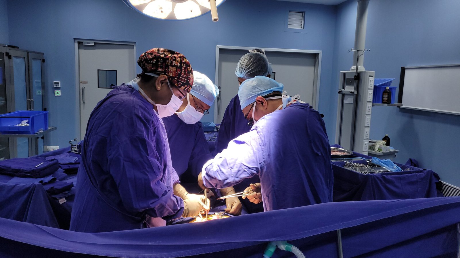
RIRS (Retrograde Intrarenal Surgery) is a procedure where a thin, flexible ureteroscope is passed through the urethra and bladder into the kidney without making any cuts on the body. A laser fibre is passed down the scope, pulverising the stone to dust in the kidney. Stones in the kidney up to about two centimetres are generally treated by RIRS. PCNL (Percutaneous Nephrolithotomy) is a procedure where a small incision is made in the back and a tract is made directly into the kidney under x-ray and ultrasound guidance. This tract is dilatated with a nephroscope which is passed through this tract to directly access and remove the stone. PCNL is indicated for larger stones (>2cm), staghorn calculi and stones in positions not accessible to flexible ureteroscopy . RIRS: quicker recovery, no skin incision PCNL: better access for larger stones.
Yes, kidney stones do come back often. Without preventive measures, approximately 50% of the patients have a recurrent stone after five years and 75% after twenty years. That is why metabolic work-up after a first stone, to identify the specific metabolic and dietary factors that are driving stone formation, is so important. Appropriate dietary modification (high fluid intake, reduced salt and animal protein, moderation of oxalate-rich foods) and medical treatment where indicated (potassium citrate for low urinary citrate, allopurinol for uric acid stones) can reduce recurrence rates by 50 percent or more. All patients with history of kidney stone should be evaluated for metabolic risk factors and provided with a personalised prevention plan.
The single most important and evidence-based preventive measure for all types of kidney stones is increased fluid intake, which alone reduces the risk of recurrence by about 50 percent. The aim is to produce at least two litres of urine a day, which means drinking about two and a half to three litres of total fluid a day in temperate climates, and more in hot conditions or during physical activity. The best practical guide is the colour of the urine, a pale straw yellow indicating adequate hydration. However, in patients with specific metabolic abnormalities (hyperparathyroidism, renal tubular acidosis, cystinuria, primary hyperoxaluria) , hydration alone may not be sufficient and targeted medical treatment beyond hydration alone is necessary.
Diet recommendations are specific to the type of stone. For calcium oxalate stones (the most common) reduce foods that are very high in oxalate (spinach, rhubarb, nuts, chocolate, strong tea, beetroot), reduce salt, moderate animal protein. Importantly, dietary calcium should not be restricted, rather adequate calcium intake (from food, not supplements) reduces oxalate absorption from the gut and reduces stone risk. For uric acid stones: cut back on foods high in purines (red meat, organ meats, shellfish, beer) and drink plenty of fluids. Salt intake should be reduced substantially in all stone types, as higher sodium intake increases urinary calcium excretion.
Recovery will depend on the procedure. After RIRS (Laser Ureteroscopy): Hospital stay 1 day; Most patients back to normal activities within 2-3 days; Full recovery in 1 week. After PCNL: 2-3 days hospital stay; 1 week light activities; 2-3 weeks full recovery. Ureteric stone removal by ureteroscopy: discharge same or next day; resume normal activities in 2-3 days. DJ stent is often left in after RIRS or ureteroscopic procedures for a period of one to three weeks, which may cause mild urinary frequency and discomfort during this period and is removed in a brief outpatient cystoscopy.
A DJ stent (double-J stent) is a soft hollow tube with one coiled end in the kidney and the other coiled end in the bladder, that is inserted inside the ureter to keep it open after endoscopic procedures of stone removal. It prevents the ureter from closing down from oedema after manipulation, allows free passage of residual stone fragments, and protects the ureter from injury while healing. In most patients a DJ stent is used for 1–3 weeks after RIRS or ureteroscopic procedures. Normal stent symptoms are frequency of urination, mild burning and at times pink urine. This is removed during a short cystoscopy as an outpatient using local anaesthetic. It is important to attend the appointment for removal of the stent as an overstayed stent can encrust with minerals and may need complex procedures to remove.
Yes, significant or longstanding kidney stone disease can impair kidney function by a number of mechanisms. An obstructing stone causes hydronephrosis , the back-pressure of accumulated urine progressively damages nephron function over days to weeks, and if obstruction is prolonged, the damage can be permanent. Recurrent infections in the context of stone disease (struvite stones, stones acting as bacterial reservoirs) lead to recurrent pyelonephritis and renal scarring. Stones in a single functioning kidney or bilateral stones are at particular risk of deterioration of renal function. This is why prompt treatment of obstructing stones and complete eradication of struvite stones is of clinical importance not only to relieve symptoms but also to preserve the kidney function .