
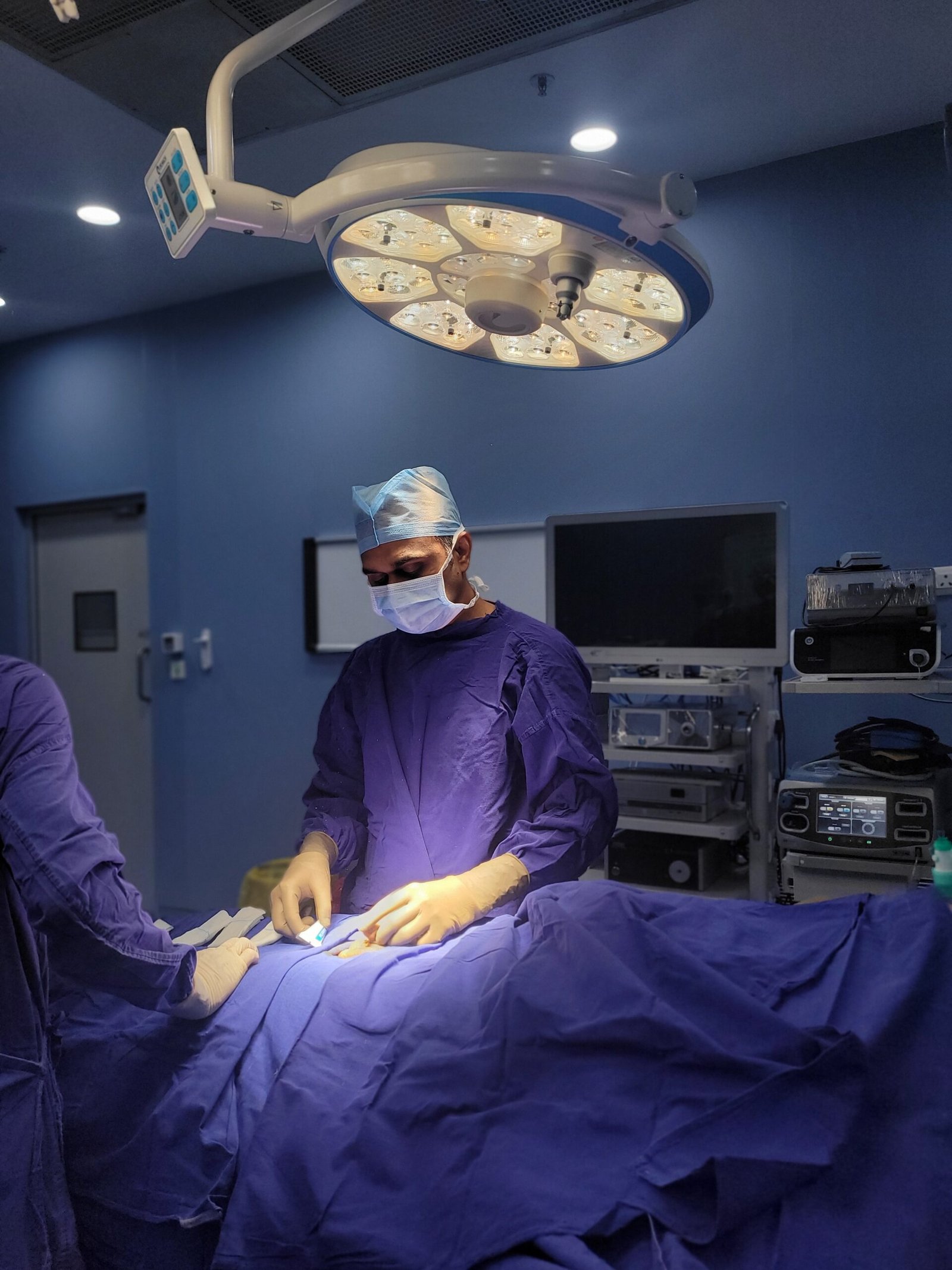
Trauma to the urethra is one of the most important injuries in urology and also one of the most time sensitive injuries. Road traffic accident, fall, medical procedure or other injuries of the urethra can cause immediate inability to urinate, long-term scarring causing narrowing of the urethral channel, and in severe cases, major complications impacting on urinary and sexual function.
Important message on urethral trauma: prompt right management is crucial. The two most important principles are: Never attempt to pass a urethral catheter without specialist guidance if urethral injury is suspected, and always obtain specialist urological care as soon as possible. Forceful catheterisation through a torn urethra in the early management can transform a manageable partial injury into a complex complete disruption.
The urethra is a tube that carries urine from the bladder to the outside of the body. In men it is about 18 to 22 cm long, from the neck of the bladder, through the prostate, through the pelvic floor and the length of the penis. In women it is much shorter , about 3 to 4 cm , from the bladder neck to just in front of the vaginal opening .
Urethral trauma is defined as any injury to this tube, be it from external force (pelvic fracture, straddle injury, penetrating wound), from medical procedures (catheterisation, cystoscopy, TURP) or from sexual trauma. The injury can be anything from a simple contusion (bruising without disruption of the structure) to a partial tear (laceration of part of the wall of the urethra) to a complete disruption (the urethra is cut through completely, and there is a gap between the two ends).
The consequences of urethral trauma are critically dependent upon the site of injury, the degree of disruption (partial versus complete) and the quality of early management. Minor injuries are often healed without surgical intervention. Major injuries, especially complete posterior urethral disruptions associated with pelvic fractures, require complex staged reconstructive surgery and have risks to long-term continence and erectile function.
Urethral trauma is classified anatomically and mechanically, these differences guide acute management and reconstructive planning:
Anterior Urethral Injury (Bulbar & Penile Urethra)
Involves the bulbar urethra (in the perineum) or the penile urethra. Most common from straddle injuries or direct trauma to the penis. Characteristic blood at meatus, perineal bruising, butterfly bruise pattern. Typically treated with suprapubic catheter drainage and urethroplasty.
Affects the membranous urethra at the level of the pelvic floor, the most vulnerable segment. Almost exclusively associated with pelvic fracture from high velocity trauma. Usually there is complete disruption. Acute suprapubic catheterisation required. Definitive posterior urethroplasty at 3-6 months.
Partial tears (involving only a portion of the urethral circumference) may heal spontaneously with catheter drainage alone. Complete disruptions (full transsection with a gap between the two ends) always require surgical reconstruction, urethroplasty, for definitive repair.
The most common mechanism is blunt trauma (straddle injury, pelvic fracture) that compresses the urethra against the pubic bones. Penetrating injuries (gunshot, stabbing) are less common but have higher rates of complex multi-organ injury requiring staged management.
Injury during catheterisation (rough insertion, or keeping the catheter balloon inflated before fully advanced) or endoscopic procedures (TURP, cystoscopy, TURBT) It creates a false passage, an iatrogenic tunnel in the periurethral tissue, which may result in stricture formation.
Knowing the mechanism of injury is important for correct classification of the urethral trauma, planning acute management and anticipating what reconstructive procedure will be needed:
Pelvic Fracture Due to Road Traffic Accident
Most common cause of posterior urethral injury High velocity impact ( motorbike, car accident ) fractures the pelvic ring. The membranous urethra tethered to the pelvic floor is sheared as the bony fragments are displaced. Significant pelvic fractures are associated with urethral injury in 10 to 25% of cases.
A fall on a hard object (bicycle handlebars, fence, scaffolding pole) compresses the bulbar urethra, the anterior most exposed portion of the urethra, against the inferior pubic rami (pubic arch). Classical mechanism of anterior urethral trauma with butterfly bruise pattern.
Most frequently caused by inflation of a Foley catheter balloon before the catheter tip is fully within the bladder, resulting in balloon inflation in the urethra, leading to immediate urethral disruption or the formation of a false passage. And also from repeated traumatic catheterizations.
Rigid cystoscopes, resectoscopes, and other endoscopic instruments can cause false passages, meatal lacerations, or urethral perforations, especially in patients with challenging anatomy or pre-existing urethral narrowing. It has been identified as a cause of iatrogenic urethral stricture.
Penile fracture (rupture of the tunica albuginea during vigorous sexual activity) is sometimes complicated by concomitant urethral injury, occurring in approximately 10 to 20% of penile fractures. Penile fracture with blood at meatus demands urgent urethral evaluation.
Urethral trauma from sexual trauma, either from assault or consensual activities involving urethral instrumentation, needs sensitive multidisciplinary management including urological care and forensic and psychological care.
Lacerations or destruction of portions of the urethra can result directly from gunshot and stab wounds to the perineum, penis, or pelvis. Complex multi-organ injuries are frequent. Management depends upon the severity of injury and haemodynamic stability of the patient.
The following signs and symptoms are suggestive of possible injury to the urethra and need immediate urological assessment before any attempt at catheterisation:
Blood at Urethral Meatus (Most Important Sign)
The most important sign of urethral injury following any pelvic trauma, straddle injury or penile trauma is blood seen at the tip of the penis (urethral meatus). Before any attempt at catheterisation, urethrogram is required without exception.
In the setting of trauma, if a mechanism for urethral injury is present and the patient is completely unable to void, a complete urethral disruption should be suspected. The appropriate emergency management is a suprapubic catheter and not a urethral catheter.
Bruising and swelling of the perineum in a characteristic “butterfly” pattern, radiating from the perineal midline outward along Colles’ fascia to both inner thighs, is pathognomonic of anterior urethral trauma due to a straddle mechanism.
Blood-stained urine following any significant trauma to the pelvis or perineum is suggestive of urethral or bladder injury. Urethrography and cystography are necessary to characterise injury prior to passing any urethral catheter.
Severe, localised perineal or pelvic pain after trauma, in particular if urination is not possible or is painful, requires urgent clinical assessment. Before attributing a musculoskeletal cause, urethral or bladder injury should be excluded.
If you are passing a catheter and feel unexpected resistance, or if you cannot advance the catheter freely into the bladder, stop immediately and call a urologist. If a catheter is introduced through an injured urethra a false passage is produced and the injury is greatly increased.
In the case of rapid swelling and discolouration (bruising, purple/black discolouration) of the scrotum or penis after trauma, extravasation of blood or urine into the fascial spaces may indicate urethral disruption. Needs urgent urological assessment.
Posted on GoogleTrustindex verifies that the original source of the review is Google. First of all Thankyou soo much sirji... Everything is all right and i am completely satisfied by sirji treatment.....one of the best urologist in indore..Posted on GoogleTrustindex verifies that the original source of the review is Google. My father is operated with prosphate, experience,nature, consultancy is very good. Allover I'm satisfied with the doctor and his services.Posted on GoogleTrustindex verifies that the original source of the review is Google. Best doctor and Best treatmentPosted on GoogleTrustindex verifies that the original source of the review is Google. Good day. Recently we visited Dr Vikas for consultation and for son's treatment at Kokilaben hospital, Indore. The hospital and its facilities are good. And Dr Vikas consultation and expert treatment were even better. He is very approachable, and listens to his patients. As parents, we are satisfied with the treatment and recommend his services. Thank you.Posted on GoogleTrustindex verifies that the original source of the review is Google. Apart from being an extremely talented urologist, Dr Vikas Singh is a great human being. I have been under his consultation for my medical conditions for last more than 2 years and I am immensely satisfied with his precision hands in surgery, his clear understanding and logical action plans to handle a complex situation of patient. I wish him all the best in his endeavours.Posted on GoogleTrustindex verifies that the original source of the review is Google. The experience is very good and nature is good while treatment
The primary imaging investigation for suspected urethral trauma is a Retrograde Urethrogram (RGU) , contrast dye is gently injected into the urethra through the external meatus and X-ray images taken as the dye flows up the urethra. This clearly shows the location, extent, and nature of any injury , whether partial or complete, anterior or posterior. RGU must be performed before any catheterisation attempt in a patient with suspected urethral injury. CT scan with urethral contrast may also be performed if available. Flexible cystoscopy may be used to directly visualise the injury in selected stable patients.
Minor partial anterior urethral contusions may heal spontaneously with suprapubic catheter drainage alone , allowing the injured urethra to rest while urine is diverted through the suprapubic catheter. However, significant partial tears and all complete urethral disruptions will not heal without leaving scar tissue , which inevitably causes urethral stricture (narrowing). Most significant urethral injuries ultimately require surgical repair (urethroplasty) to achieve a satisfactory, durable result. The question is not usually if surgery is needed, but when and what type.
Endoscopic urethrotomy (OIU , optical internal urethrotomy) is a minimally invasive endoscopic procedure where a laser or knife is used to cut through urethral scar tissue from within the urethra , performed without any external incision. It is effective for short, thin strictures but has a high long-term recurrence rate (50 to 70%) and is generally not appropriate for complex traumatic strictures or complete posterior urethral disruptions. Urethroplasty is an open reconstructive procedure that excises the damaged segment and either rejoins the healthy ends (anastomotic urethroplasty) or inserts a tissue graft (substitution urethroplasty). Urethroplasty has long-term success rates of 85 to 95% and is the definitive treatment for significant traumatic urethral injuries.
Posterior urethral injuries , particularly those involving the membranous urethra at the pelvic floor , are in close proximity to the neurovascular bundles responsible for erectile function. Erectile dysfunction after posterior urethral trauma from pelvic fracture affects approximately 30 to 40% of patients. This may result from direct nerve injury at the time of trauma, from the pelvic fracture disrupting penile blood supply, or from the reconstructive surgery. Dr. Vikas Singh performs nerve-sparing posterior urethroplasty wherever oncologically and anatomically feasible, and provides dedicated assessment and management of post-traumatic erectile dysfunction as part of the long-term follow-up programme.
After urethroplasty, a urethral catheter is typically in place for 3 to 4 weeks , allowing the reconstruction to heal around it. Most patients are discharged 3 to 5 days after surgery. After catheter removal, a voiding cystourethrogram (VCUG) or uroflowmetry is performed to confirm successful voiding through the repaired urethra. Most patients return to light work within 3 to 4 weeks and full activity within 6 to 8 weeks. Regular follow-up uroflowmetry at 3, 6, and 12 months detects any early recurrent stricture formation , which, if caught early, is more easily managed.