

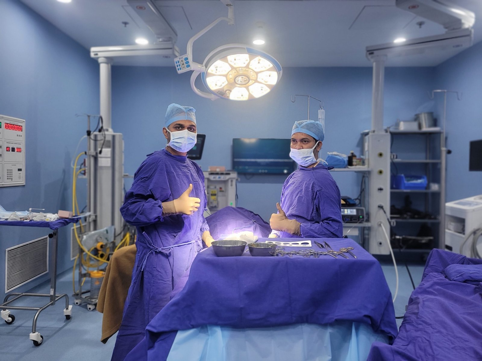
The urinary bladder is one of those organs that most of us take for granted, until it stops working properly. A healthy bladder fills silently, comfortably holds urine, and then empties completely on demand. If something goes wrong – a burning infection, a stone that causes pain, a tumour discovered on a scan, the humiliation of leaking urine at an unguarded moment – the impact on daily life is immediate, profound and often very personal.
Bladder problems include the very common (urinary tract infections, overactive bladder), the potentially serious (bladder cancer, vesicovaginal fistula), and the chronically misunderstood (interstitial cystitis, bladder neck obstruction). What they have in common is the need for a proper specialist assessment, because many bladder symptoms overlap, and the correct diagnosis is the essential first step to effective treatment.
Bladder Cancer & Bladder Tumours
Bladder cancer is the most common malignancy of the urinary tract. It presents most commonly with painless haematuria. Most are transitional cell carcinomas of the lining of the bladder. TURBT is used to treat early stage tumours. Cystectomy or radiation and chemotherapy is used for muscle invasive disease.
Bladder stones are mineral deposits that form when urine sits in the bladder . The most common causes for this are BPH , bladder outflow obstruction , or foreign bodies . They cause pain, haematuria and interrupted stream of urine. Treatment : For large stones , cystoscopic laser fragmentation or open cystolithotomy .
Cystitis is an inflammation of the bladder lining, most commonly due to a bacterial infection (UTI), and is characterized by burning urination, frequency, urgency and pelvic discomfort. More common in women, because of shorter urethra. Treatment: appropriate antibiotics. Recurrent cases: investigate for underlying causes.
Bladder neck obstruction is a functional or anatomic narrowing at the bladder outlet leading to difficulty voiding, weak stream and incomplete emptying. Common in younger men without BPH. Urodynamic diagnosis. Alpha-blockers or bladder neck incision (BNI) endoscopically.
Interstitial cystitis (IC) is a chronic bladder condition that causes chronic pelvic pain, pressure, severe urinary urgency and frequency, often without infection. The lining of the bladder is inflamed and overly sensitive. Management is multimodal: dietary modification, bladder instillations, medications and pelvic physiotherapy.
Urinary incontinence is the involuntary loss of urine and is experienced by millions of people around the world. It can have a major impact on quality of life. Types: Stress incontinence (leakage with coughing / sneezing) Urgency incontinence (overactive bladder) Mixed . They were given pelvic floor therapy, medication or surgery.
VVF is an unnatural channel between the bladder and vagina that causes the continuous leakage of urine through the vagina. It is most commonly due to obstetric trauma in a difficult labour or after gynaecological surgery. Surgical repair (open or laparoscopic) is required. Good results can be achieved in experienced hands.
Bladder symptoms are often embarrassing, so they’re under-reported and under-treated. Every sign below deserves medical attention, as effective treatment exists for each.
Blood in Urine (Haematuria)
Any bladder symptom that is persistent, recurring, or involves blood in the urine should be assessed by a specialist. These situations need prompt or urgent referral.
Bladder symptoms are often embarrassing and are therefore often under-reported and under-treated. Any of the following warning signs should be taken seriously as effective treatment exists for each and each warrants medical assessment.
Blood in Urine (Haematuria) – Painless or Painful
Refer any bladder symptom that is persistent, recurrent or has blood in the urine for specialist assessment. Examples of situations requiring prompt or urgent urological referral include:
Blood in Urine – Even Once Should Never Be Ignored
Accurate diagnosis of bladder disorders requires a combination of clinical evaluation, targeted laboratory investigations and imaging or endoscopic studies. Dr. Vikas Singh follows a systematic, evidence based diagnostic approach for each and every patient:
Urine Routine & Culture | Infection, blood, protein; identifies causative bacteria | Cystitis; recurrent UTI; haematuria screen |
Urine Cytology | Abnormal (malignant) cells shed from bladder lining | Bladder cancer screening; high-grade tumour detection |
Ultrasound KUB | Bladder wall thickness, mass, stone, post-void residual | Initial assessment of most bladder conditions |
CT Urogram | Entire urinary tract , tumour, stone, structural abnormality | Bladder cancer staging; haematuria investigation |
Cystoscopy | Direct visual examination of bladder interior and urethra | Definitive bladder cancer diagnosis; unexplained haematuria |
Urodynamic Studies | Bladder capacity, compliance, detrusor function, outlet resistance | Incontinence; voiding dysfunction; neurogenic bladder |
MRI Pelvis | Bladder wall invasion depth; lymph node involvement; VVF tract | Muscle-invasive bladder cancer staging; VVF planning |
Retrograde/Fistulogram | Demonstrates fistula tract with contrast | VVF confirmation; complex fistula assessment |
Urine Routine Microscopy & Urine Culture
These are the first tests for any urinary symptom. A dipstick quickly shows signs of infection or blood, and microscopy confirms red cells, white cells, and bacteria. Culture identifies the exact organism and the antibiotics that work best, which matters most for recurrent or resistant infections. A clean mid-stream sample is used to avoid contamination.
Urine Cytology for Bladder Cancer Screening
Urine cytology checks cells shed from the bladder lining for cancerous changes. It works best for high-grade bladder cancer and is less reliable for low-grade tumors. It’s used when investigating blood in urine, monitoring after treatment, and screening high-risk workers in the dye, rubber, and chemical industries. A positive result makes cystoscopy more urgent.
Ultrasound KUB for Bladder Mass, Stone & Wall Thickening
A KUB ultrasound is the simplest and most useful first scan for bladder problems. It can show bladder growths, stones, wall thickening, leftover urine after voiding, and kidney swelling caused by blockage. It’s safe, radiation-free, and should be the first scan for urinary symptoms or unexplained blood in urine.
CT Urogram for Comprehensive Bladder & Upper Tract Evaluation
A CT urogram is a contrast scan that images the entire urinary tract. It’s the definitive test for blood in urine and the standard scan for staging bladder cancer. It shows the bladder, kidneys, and ureters, and can detect upper tract tumors, swollen lymph nodes, and spread to other organs, which ultrasound can’t.
Cystoscopy: Direct Visualisation Inside the Bladder
Cystoscopy is the best test for bladder disease. A thin telescope passed through the urethra lets the doctor see tumors, inflammation, stones, and structural problems directly. Done in the clinic under local anesthetic gel, it takes about five minutes, and no scan can replace it for unexplained blood in urine. If a tumor is found, it can be removed and tested during a TURBT under anesthesia.
Urodynamic Studies for Bladder Function Assessment
Urodynamics measures how well the bladder and urethra work, something no scan can show. It records bladder capacity, stretch, urges, and pressure during filling and emptying, helping tell blockage apart from a weak bladder. It’s the gold standard test before surgery for incontinence, voiding problems, or neurogenic bladder.
MRI Pelvis for Bladder Cancer Staging & VVF Evaluation
Pelvic MRI gives the most detailed view of the bladder wall and nearby tissues. In muscle-invasive bladder cancer, it shows how deep the tumor has grown and whether it has spread to nearby fat or lymph nodes, which guides treatment choices. For VVF, it maps the fistula tract and surrounding anatomy to plan surgery.
Retrograde Pyelogram & Fistulogram When Required
When a CT urogram isn’t enough, a retrograde pyelogram injects contrast dye up the ureter to show it and the kidney’s drainage system in detail. A fistulogram injects dye into a fistula tract to reveal its path and connections, giving a roadmap for repair. Both are done during cystoscopy under anesthesia, with X-ray guidance.
Your bladder health depends a lot on daily habits. Many bladder problems can be prevented or easily managed with simple, evidence-based steps.
How Much Water Should You Drink for Bladder Health
Foods & Drinks That Irritate the Bladder
Pelvic Floor Exercises for Bladder Control
How to Prevent Recurrent Bladder Infections
Smoking & Bladder Cancer: A Direct Connection
Regular Screening for High-Risk Bladder Cancer Patients
Bladder conditions span the full spectrum of urological complexity , from common infections to serious cancers to rare conditions like VVF. Expert management requires both broad clinical knowledge and specific surgical skills. Here is why patients across Indore and Central India trust Dr. Vikas Singh for bladder care:
As a trained Genito-Uro Oncologist, Dr. Vikas Singh brings specialist cancer expertise to bladder tumour diagnosis and management , from TURBT for initial diagnosis and staging to advanced surveillance protocols and decisions about radical cystectomy for muscle-invasive disease. Bladder cancer patients receive oncologically expert, evidence-based care from a specialist , not a generalist.
Comprehensive Diagnostic Capability
Dr. Vikas Singh offers the full range of bladder investigations , flexible and rigid cystoscopy, urodynamic studies, CT urogram, MRI pelvis, urine cytology, and retrograde pyelogram , all available at Vishesh Jupiter Hospital, Indore. Patients do not need to travel to multiple facilities for different tests.
Full Surgical Spectrum , Endoscopic to Open
From TURBT for bladder tumours and cystolithotripsy for bladder stones to open VVF repair, bladder neck incision, and complex bladder reconstruction , Dr. Vikas Singh performs the complete range of bladder surgical procedures, matching the approach to each patient’s condition and clinical needs.
Women’s Bladder Health & VVF Expertise
Dr. Vikas Singh provides dedicated care for women’s bladder conditions , including urinary incontinence (urodynamic assessment and surgical correction), interstitial cystitis management, and vesicovaginal fistula repair. VVF repair requires specialist surgical experience , Dr. Vikas Singh has specific expertise in this challenging and life-changing procedure.
15+ Years of Experience & 10,000+ Procedures
With over 15 years of focused urological practice encompassing thousands of bladder procedures and a specialist background in genito-urinary oncology, Dr. Vikas Singh brings the clinical depth that bladder disease , in all its variety , demands.
Compassionate Care for Sensitive Conditions
Bladder problems , particularly incontinence, VVF, and painful bladder syndrome , carry significant emotional and social burden. Dr. Vikas Singh provides a confidential, non-judgmental consultation environment where patients feel safe discussing conditions they may have been managing alone for years.
Vishesh Jupiter Hospital, Indore
All bladder investigations and procedures are performed in a modern, fully equipped hospital with dedicated endoscopy and surgical facilities, advanced imaging, urodynamics laboratory, and integrated oncology services , providing world-class bladder care right here in Indore.
Urine Routine Microscopy & Urine Culture
Urine dipstick test, microscopy and culture are the first line investigations in any patient presenting with urinary symptoms. A dipstick test for blood, protein, leucocytes and nitrites will indicate immediately if infection or haematuria is likely. Microscopy: Confirms the presence of red blood cells, white blood cells and bacteria under the microscope. Culture will tell us the specific organism that is causing the infection and antibiotic sensitivity profile which is important to decide the most effective treatment especially in case of recurrent or resistant infections. The usual type of specimen is a mid-stream urine (MSU) sample, collected cleanly to avoid contamination by bacteria on the external skin.
Urine Cytology for Bladder Cancer Screening
Urine cytology looks for abnormal (malignant) features in cells shed from the bladder lining into the urine. It is most sensitive for high-grade urothelial carcinoma, the most aggressive form of bladder cancer, which reliably sheds cytologically abnormal cells. It is less sensitive for low-grade tumours that shed fewer abnormal cells. Urine cytology is used in the haematuria investigation protocol, in surveillance after treatment of bladder cancer and in occupational screening programmes for high risk workers (dye, rubber and chemical industry workers). A positive cytology in a patient with haematuria increases the urgency of cystoscopy significantly.
Ultrasound KUB for Bladder Mass, Stone & Wall Thickening
A kidney, ureter, bladder (KUB) ultrasound is the most simple, most available and most informative first imaging study for most bladder conditions. It identifies bladder masses (polypoid lesions projecting into the bladder lumen), bladder stones (dense echogenic deposits with acoustic shadowing), focal or diffuse bladder wall thickening (suggestive of tumour, cystitis or neurogenic changes), post void residual urine volume and upper tract dilation suggesting bladder outlet obstruction affecting the kidneys. Ultrasound is safe, radiation free and available as an outpatient investigation it should be the first imaging test in any patient with lower urinary tract symptoms or unexplained haematuria.
CT Urogram for Comprehensive Bladder & Upper Tract Evaluation
CT urography (CTU) is a three-phase computed tomography scan with intravenous contrast, which images the entire urinary tract. It is the definitive radiological investigation for haematuria and the standard staging investigation for bladder cancer. It shows the bladder in detail, the upper urinary tract (kidneys and ureters), any other upper tract tumours (which are present in about three percent of patients with bladder tumour), lymphadenopathy and distant metastases. CTU offers information not available with ultrasound alone, especially for ureteral tumours, upper tract anatomy and lymph node involvement.
Cystoscopy – Direct Visualisation Inside the Bladder
The test of choice for diseases of the bladder is cystoscopy. A thin telescope, either flexible or rigid, is passed through the urethra into the bladder, and the interior of the bladder can be seen directly. The surgeon, using cystoscopic vision, can identify bladder tumours (confirming their size, location, number and appearance), evidence of cystitis or inflammation, bladder stones, abnormal ureteric orifices suggestive of VUR, bladder neck configuration and any structural abnormalities. Flexible cystoscopy in the outpatient setting under local anaesthetic gel is the definitive investigation for unexplained haematuria. It takes about five minutes, and ultrasound or CT alone cannot substitute for this. On diagnosis of a tumour rigid cystoscopy under anaesthesia (TURBT) is performed for resection and definitive histological diagnosis simultaneously.
Urodynamic Studies for Bladder Function Assessment
Urodynamics is a specialized test that provides objective characterization of bladder and urethral function and provides information that no imaging study can. A filling cystometry study measures bladder capacity, compliance (ability to stretch without a pressure rise), sensations during filling, and the presence of involuntary detrusor contractions (overactive bladder). A pressure-flow voiding study simultaneously records the contractile pressure of the bladder during voiding and the urine flow rate, which provides a means for calculation of the bladder outlet resistance and distinction between obstruction and detrusor weakness as the cause of poor flow. Urodynamics is the gold standard test prior to surgery for incontinence, voiding dysfunction or neurogenic bladder.
MRI Pelvis for Bladder Cancer Staging & VVF Evaluation
MRI of the pelvis is the most detailed imaging of soft tissue of the bladder wall, adjacent structures, lymph nodes and pelvic anatomy. In muscle-invasive bladder cancer MRI accurately defines the depth of tumour invasion into the bladder wall, whether tumour has spread through the wall into perivesical fat and pelvic lymph node involvement, information directly guiding the choice between neoadjuvant chemotherapy followed by cystectomy, bladder-preserving chemoradiation and palliative treatment. MRI plays an important role in the evaluation of VVF by depicting the fistula tract, its relation to the bladder trigone and ureteric orifices, associated tissue injury and anatomy of surrounding structures; all of which are important for surgical planning.
Retrograde Pyelogram & Fistulogram When Required
If a CT urogram does not give enough information or if the ureteric anatomy needs to be precisely characterized before surgery, a retrograde pyelogram—in which contrast dye is injected up the ureter through a cystoscopically placed catheter—provides detailed imaging of the ureter and renal collecting system. A fistulogram, the injection of contrast directly into a fistula tract, outlines the tract’s course, origin, and relationship to adjacent structures, and provides a roadmap for surgical repair. Both investigations are carried out during a cystoscopic examination under anaesthesia, under fluoroscopic guidance.
The health of our bladder is very much affected by how we live our lives. There are many bladder conditions that can be prevented or are very manageable with simple daily habits. This is evidence-based advice on keeping your bladder in good health:
How Much Water Should You Drink for Bladder Health
Foods & Drinks That Irritate the Bladder
Pelvic Floor Exercises for Bladder Control
How to Prevent Recurrent Bladder Infections
Smoking & Bladder Cancer – A Direct Connection
Regular Screening for High-Risk Bladder Cancer Patients
Bladder conditions span the full spectrum of urological complexity , from common infections to serious cancers to rare conditions like VVF. Expert management requires both broad clinical knowledge and specific surgical skills. Here is why patients across Indore and Central India trust Dr. Vikas Singh for bladder care:
Posted on GoogleTrustindex verifies that the original source of the review is Google. One of the best urologist i have even met. Thank you Dr. VikasPosted on GoogleTrustindex verifies that the original source of the review is Google. Mene Dr vikas singh sir ko jupiter hospital me prostate ke liye dikhaya tha, dr vikas sir ne bahut ache se bataya thanks dr sirPosted on GoogleTrustindex verifies that the original source of the review is Google. Dr. Vikas Singh is undoubtedly one of the finest urologists in Indore. Along with his excellent medical expertise, what truly sets him apart is his kindness, humility, and genuine understanding of the difficulties faced by patients. Instead of being rigid in his approach, he always tries to find the best possible solution in the patient's interest, making them feel heard, understood, and cared for. His compassionate nature and patient-first attitude make him a truly exceptional doctor. I feel blessed and grateful to have received my treatment under his supervision. May God always bless him with good health, happiness, and success, and give him the strength to continue serving humanity with the same dedication and compassion. 🙏Posted on GoogleTrustindex verifies that the original source of the review is Google. Dr vikas sir jupiter hospital me mere father ko dikhya, sir bahut ache se treat Kiya or sir ka knowlege bhi bahut cha hePosted on GoogleTrustindex verifies that the original source of the review is Google. First of all Thankyou soo much sirji... Everything is all right and i am completely satisfied by sirji treatment.....one of the best urologist in indore..Posted on GoogleTrustindex verifies that the original source of the review is Google. My father is operated with prosphate, experience,nature, consultancy is very good. Allover I'm satisfied with the doctor and his services.Posted on GoogleTrustindex verifies that the original source of the review is Google. Best doctor and Best treatment
Painless visible haematuria , blood in the urine without any associated pain , is the most important and most common presenting symptom of bladder cancer. It occurs in approximately 85 percent of bladder cancer patients at some point in their disease course and is classically intermittent , it may appear once, disappear for weeks or months, and then recur. This intermittent nature is dangerous because patients often assume the episode was benign when it resolved without treatment. Any single episode of painless visible haematuria in an adult over 40 should trigger urgent urological assessment including cystoscopy and CT urogram , regardless of whether it has occurred only once or has resolved spontaneously.
An uncomplicated lower urinary tract infection (cystitis) in an otherwise healthy young woman is a common, straightforward condition that responds well to a short course of appropriate antibiotics and is not considered serious. However, a UTI that extends to the kidney (pyelonephritis) , producing fever, rigors, and loin pain , is a more serious upper urinary tract infection requiring more intensive treatment. UTIs in men are always clinically significant and require investigation for underlying structural causes (BPH, bladder stone, stricture). Recurrent UTIs in any patient , male or female , warrant investigation to identify and treat any predisposing anatomical or functional abnormality.
Interstitial cystitis (IC) , also called painful bladder syndrome , is a chronic condition characterised by persistent pelvic pain or pressure, severe urgency and frequency, and pain that worsens as the bladder fills and partially relieves after voiding , in the absence of proven infection. The condition involves dysfunction of the bladder’s protective mucus lining, allowing urinary irritants to penetrate and trigger nerve hypersensitivity. IC is a condition that is managed rather than cured for most patients , but management can achieve very significant symptom control. Treatment includes dietary modification (eliminating bladder irritants), bladder instillations (with heparin, lignocaine, or DMSO), oral medications (amitriptyline, hydroxyzine, pentosan polysulphate), and pelvic floor physiotherapy. Many patients achieve substantial relief with a tailored multimodal approach.
A vesicovaginal fistula (VVF) is an abnormal tract or opening between the bladder and the vagina, causing continuous or intermittent leakage of urine through the vagina. The most common causes are difficult prolonged labour (obstructed labour, particularly common in settings with limited access to emergency obstetric care), gynaecological surgery (particularly hysterectomy), and pelvic radiotherapy. The diagnosis is confirmed by clinical examination, dye test (instilling methylene blue into the bladder and checking for vaginal staining), cystoscopy, and MRI pelvis. Treatment is surgical repair , performed either open (through the vagina or abdomen) or laparoscopically. Success rates with experienced surgeons exceed 90 percent, and most women are completely cured of their leakage after a single repair.
Cystoscopy is the direct visual examination of the bladder interior using a thin telescope (cystoscope) passed through the urethra. Flexible cystoscopy , performed in the outpatient clinic under local anaesthetic lubricant gel , is the standard approach for diagnostic cystoscopy in adults. It takes approximately five minutes and causes mild to moderate urethral discomfort that most patients tolerate without difficulty. It is not the severely painful procedure many patients fear. Rigid cystoscopy under general or spinal anaesthesia is used when a procedure needs to be performed at the same time , such as tumour resection (TURBT), biopsy, or stone fragmentation. Cystoscopy is the only investigation that can directly visualise the bladder interior and identify early bladder tumours that ultrasound and CT may miss.
Yes , urinary incontinence has highly effective treatments for all types. Stress urinary incontinence (leakage with physical exertion) responds very well to pelvic floor muscle training in mild to moderate cases (achieving dryness or significant improvement in 60 to 70 percent of patients with supervised physiotherapy) and to surgical procedures such as mid-urethral sling in more severe or refractory cases (success rates of 85 to 90 percent at five years). Urgency incontinence (overactive bladder) is treated with bladder training, lifestyle modification, antimuscarinic or beta-3 agonist medications, botulinum toxin bladder injection for refractory cases, and sacral neuromodulation for severe drug-resistant cases. Mixed incontinence is addressed through a combination of approaches targeting both components.
Recurrent UTIs (two or more per six months or three or more per year) require investigation to identify any underlying predisposing factors. In women, common contributing factors include sexual activity patterns, contraceptive method (spermicides increase risk), post-menopausal vaginal atrophy (oestrogen deficiency impairs mucosal defences), bladder prolapse causing incomplete emptying, and diabetes. In men, any UTI should prompt investigation for BPH, bladder outlet obstruction, bladder stone, or urinary stasis. In both sexes, bladder stones, bladder tumours, incomplete bladder emptying (elevated post-void residual), vesicoureteral reflux, and immunocompromise are important structural causes that must be excluded by ultrasound, urine cultures, and cystoscopy before attributing recurrent UTIs to simple bad luck.
Bladder cancer is staged by the depth of tumour invasion into the bladder wall, using the TNM classification. Non-muscle-invasive bladder cancer (NMIBC) , confined to the bladder lining (Ta, T1, CIS) , is the most common presentation and is treated by TURBT with or without intravesical instillations (BCG or mitomycin C into the bladder) followed by cystoscopic surveillance. Muscle-invasive bladder cancer (MIBC, T2 and above) , where the tumour has invaded the bladder muscle , requires more aggressive treatment: radical cystectomy (surgical removal of the bladder and creation of a urinary diversion) with or without neoadjuvant chemotherapy, or bladder-preserving concurrent chemoradiation for selected patients. Staging by CT urogram and MRI pelvis determines local extent and lymph node involvement before treatment decisions are finalised.
TURBT stands for Transurethral Resection of a Bladder Tumour , it is the primary surgical procedure for treating and diagnosing bladder cancer. Using a rigid cystoscope passed through the urethra under general or spinal anaesthesia, the surgeon uses an electrically activated resecting loop to shave away the tumour from the bladder wall in fragments, which are sent for histological analysis. TURBT simultaneously diagnoses the cancer (histology, grade) and treats it , removing all visible tumour tissue. For non-muscle-invasive bladder cancer (tumour confined to the bladder lining and not invading the muscle wall), TURBT followed by surveillance cystoscopy is the standard curative treatment. For muscle-invasive bladder cancer (tumour invading the bladder muscle), TURBT establishes the diagnosis and staging, and is followed by radical cystectomy or bladder-preserving chemoradiation.