
PUJO , Pelvic Ureteric Junction Obstruction , is one of the most common causes of significant kidney swelling ( hydronephrosis ) in Children as well as Adults . It is a condition in which the junction between the collecting system of the kidney (renal pelvis) and the ureter becomes abnormally narrowed or functionally obstructed, preventing urine from draining freely from the kidney into the ureter.
PUJO is usually an incidental finding on a fetal prenatal ultrasound, on a scan of the abdomen for another reason in an adult, or in the workup of recurrent flank pain, kidney infections, or declining kidney function. It may become large and cause progressive damage to the kidney, which can be completely avoided by timely surgical repair.
The renal pelvis is a funnel-shaped collecting space within the centre of the kidney. Urine collects in the renal pelvis after being produced by the kidney’s filtering units (nephrons). Urine flows down from the renal pelvis into the ureter, the tube that conducts urine to the bladder. The Pelvi-Ureteric Junction (PUJ) is where the renal pelvis becomes the ureter.
In PUJO, this junction is abnormally narrow, kinked or functionally ineffective, so that there is no free drainage of urine from the renal pelvis into the ureter. The kidney still produces urine that collects in the renal pelvis but cannot flow through the obstructed PUJ at a fast enough rate. As the pressure increases, the pelvis expands and the kidney inflates – a condition called hydronephrosis (from the Greek hydro = water, nephrosis = kidney condition).
PUJO is the most common cause of antenatal hydronephrosis and the most frequent diagnosis when a fetal kidney is seen to be dilated on a prenatal ultrasound scan. It can also occur in adults, either as a congenital anomaly that has been previously compensated but now becomes clinically evident or as an acquired anomaly due to stone, infection, surgery or external compression. Untreated significant PUJO results in progressive injury of the functional tissue of the kidney due to sustained back-pressure damage.
Understanding the normal anatomy and function of the PUJ explains why obstruction at this particular point is so clinically important:
Anatomy of Kidney Pelvis & Ureter Connection
The renal pelvis is a funnel-shaped expansion in the centre of the kidney. It gradually tapers to become the proximal ureter at the PUJ. The narrowest exit point of the funnel is the PUJ.
Urine flows from the calyces into the renal pelvis by gravity and peristaltic (wave-like) contractions of the pelvic wall. Peristaltic waves at the PUJ drive urine into the upper ureter and downwards to the bladder.
Obstruction at the PUJ prevents urine from passing into the ureter as quickly as it is made, causing back-pressure to build up in the renal pelvis. As urine pools, the pelvis dilates progressively.
Sustained back-pressure in the dilated renal pelvis is transmitted to the nephrons , the filtering units of the kidney , squeezing them and gradually impairing their filtration function , causing irreversible kidney damage if untreated.
PUJO can present dramatically different depending on the severity of the obstruction, patient’s age and how long the condition has been present. Some patients are asymptomatic, others present with severe recurrent pain:
Intermittent Flank Pain & Back Pain (Dietl's Crisis)
The symptoms of symptomatic PUJO in the adult are episodes of severe, cramping flank pain, especially following the consumption of large quantities of fluid. These episodes are known as Dietl’s crisis and occur when there is increased urine production that overwhelms the obstructed PUJ.
The severity of PUJO-related flank pain often triggers nausea and vomiting . This may result in patients presenting to emergency departments with a picture similar to acute ureteric colic from a kidney stone.
The dilated renal pelvis with its stagnant urine is a perfect medium for bacterial colonisation. Recurrent febrile UTIs, especially pyelonephritis, in a patient with known hydronephrosis should raise suspicion for PUJO and prompt further evaluation.
The dilated obstructed renal pelvis in PUJO patients is more prone to minor trauma and trivial activities like exercise can induce mucosal bleeding and haematuria. Any haematuria in a patient known to have hydronephrosis warrants investigation.
In infants and young children presenting with severe PUJO, the massively dilated kidney may be palpable as a smooth, non-tender mass in the flank or upper abdomen . Occasionally, the abdominal mass is the presenting symptom that brings the child to medical attention.
Secondary hypertension due to chronic renal obstruction with activation of the renin–angiotensin system. New or worsening hypertension in a patient with diagnosed hydronephrosis could indicate clinically significant PUJO and should be treated.
The most objective evidence of progressive renal damage with PUJO that warrants surgical repair is a rising creatinine or falling eGFR , or worsening differential kidney function on MAG3 renography, in a patient with hydronephrosis.
PUJO may be congenital (present at birth) or acquired (developing later in life). The most appropriate surgical approach is dictated by the underlying cause:
Abnormal Muscle Development at PUJ (Congenital)
The renal pelvis is a funnel-shaped expansion in the centre of the kidney. It gradually tapers to become the proximal ureter at the PUJ. The narrowest exit point of the funnel is the PUJ.
An accessory lower pole renal artery or vein crosses the ureter at the PUJ with an external kink and intermittent or sustained compression. This is an important etiology in adults presenting with symptoms of intermittent PUJO.
PUJ obstruction and hydronephrosis secondary to stone impaction. This is a secondary, potentially reversible obstruction, treatable with the stone, unlike intrinsic PUJO. Ultrasound and CT differentiate these from primary PUJO.
Secondary stenosis (acquired) Fibrotic scarring at the PUJ from prior kidney surgery, ureteral injury or trauma. Recurrent PUJO in previously treated patients can be caused by post-pyeloplasty restenosis.
A fixed, fibrous narrowing at the level of the PUJ occurs as a result of ureteral stricture from previous infection, instrumentation or ischaemia. Some short selected strictures may be amenable to endoscopic endopyelotomy (incision of the PUJ from within).
Secondary hydronephrosis can occur from compression of the PUJ from outside, due to retroperitoneal tumours, enlarged lymph nodes or masses in the vicinity of the kidney. Imaging finds the compressing mass, treatment targets the underlying cause.
The Society for Fetal Urology ( SFU ) grading system is used to grade the severity of hydronephrosis from PUJO and is used for both prenatal and postnatal assessment or the analogous grading system for adults is used . The urgency of the treatment is directly related to the grade:
Grade | Ultrasound Appearance | Kidney Function Impact | Management Approach |
Grade 1 , Mild | Slight dilatation of renal pelvis only. Calyces normal. | Minimal , kidney function generally preserved | Watchful waiting + 6-monthly ultrasound; most resolve spontaneously in children |
Grade 2 , Moderate | Renal pelvis dilated. Calyces mildly dilated but cortex normal thickness. | Mild , kidney function usually preserved | Close surveillance with MAG3 renogram; surgical intervention if function declines or symptoms develop |
Grade 3 , Severe | Renal pelvis and calyces significantly dilated. Cortex beginning to thin. | Moderate , kidney function may be reduced on renogram | Pyeloplasty usually recommended , risk of progressive function loss without surgery |
Grade 4 , End Stage | Gross dilatation of pelvis and calyces. Cortex severely thinned. | Significant , differential function often below 20–25% | Urgent pyeloplasty if residual function remains; nephrectomy considered if function non-recoverable |
Grade 1 & 2 , Surveillance: The majority of children with prenatally diagnosed Grade 1 and 2 hydronephrosis do not require surgery. They are followed with watchful waiting, serial ultrasounds at 3-6 monthly intervals and a MAG3 renal function scan at 4-6 weeks of age. Most Grade 1 and many Grade 2 cases spontaneously resolve during the first 18 to 24 months of life. Antibiotic prophylaxis can be given for the prevention of UTI during surveillance.
Grade 3 & 4 , Pyeloplasty: The main indication for pyeloplasty is grade 3 hydronephrosis with evidence of obstruction on MAG3 renography (differential function < 40%, obstruction pattern or falling function on sequential scans). Grade 4 hydronephrosis requires urgent evaluation . If the kidney has over 20 to 25% differential function , pyeloplasty provides the best opportunity for partial functional recovery . If function is minimal or absent, consider nephrectomy.
Posted on GoogleTrustindex verifies that the original source of the review is Google. First of all Thankyou soo much sirji... Everything is all right and i am completely satisfied by sirji treatment.....one of the best urologist in indore..Posted on GoogleTrustindex verifies that the original source of the review is Google. My father is operated with prosphate, experience,nature, consultancy is very good. Allover I'm satisfied with the doctor and his services.Posted on GoogleTrustindex verifies that the original source of the review is Google. Best doctor and Best treatmentPosted on GoogleTrustindex verifies that the original source of the review is Google. Good day. Recently we visited Dr Vikas for consultation and for son's treatment at Kokilaben hospital, Indore. The hospital and its facilities are good. And Dr Vikas consultation and expert treatment were even better. He is very approachable, and listens to his patients. As parents, we are satisfied with the treatment and recommend his services. Thank you.Posted on GoogleTrustindex verifies that the original source of the review is Google. Apart from being an extremely talented urologist, Dr Vikas Singh is a great human being. I have been under his consultation for my medical conditions for last more than 2 years and I am immensely satisfied with his precision hands in surgery, his clear understanding and logical action plans to handle a complex situation of patient. I wish him all the best in his endeavours.Posted on GoogleTrustindex verifies that the original source of the review is Google. The experience is very good and nature is good while treatment
No. PUJO and kidney stones are two different entities, although both can present with flank pain and hydronephrosis . A kidney stone is a solid mass of minerals that forms in the kidney and can block the PUJ if it becomes lodged there. PUJO is a structural narrowing or functional blockage of the PUJ itself. Usually congenital (present from birth) or from crossing vessels, scarring or stricture. The key diagnostic distinction: CT urogram (which visualises the anatomy and any stones) and MAG3 renography (which assesses drainage and function) are the investigations which clearly separate the two.
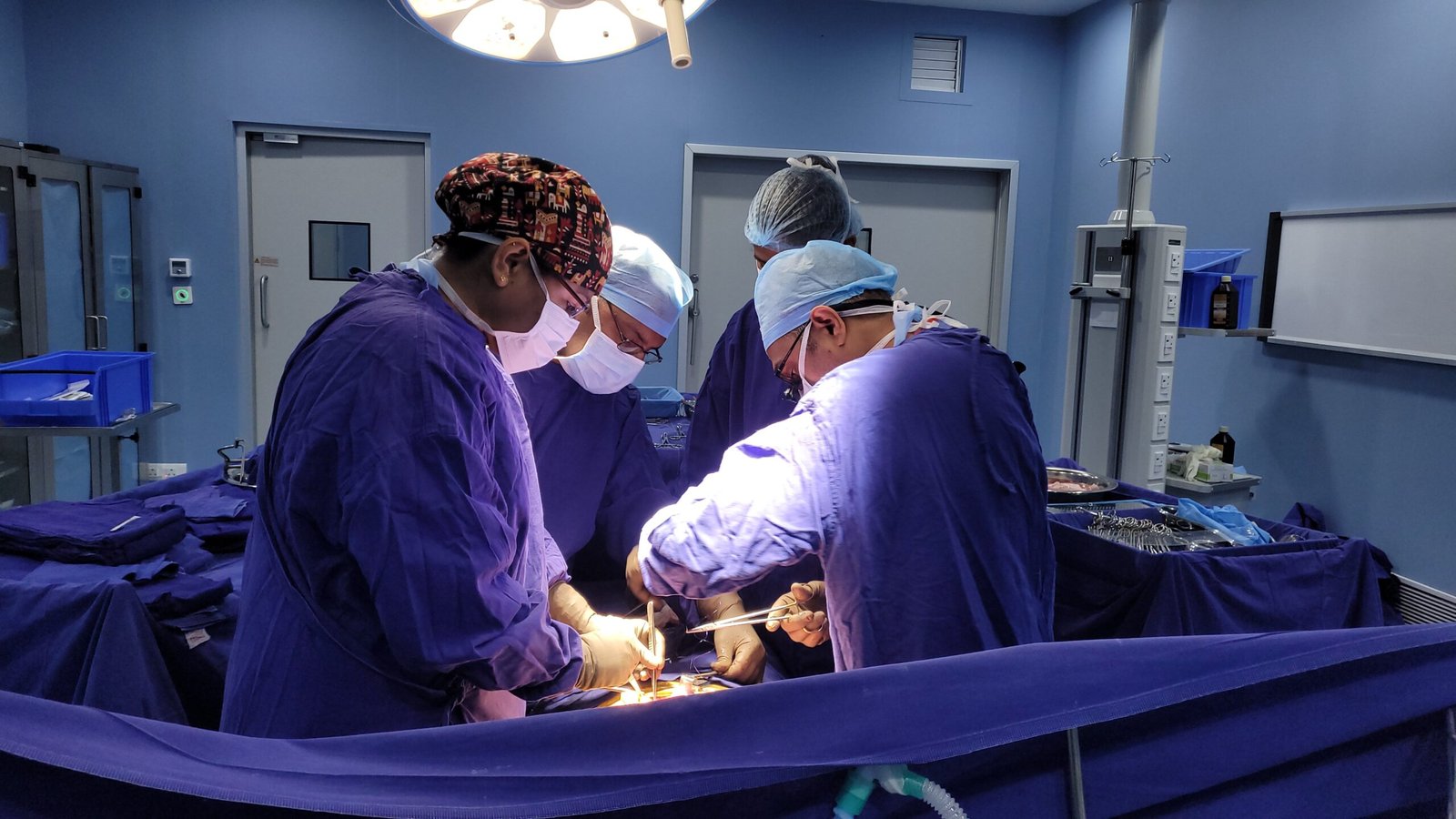
Laparoscopic pyeloplasty is the surgical gold standard for treating PUJO. Through 3 to 4 small keyhole incisions (5 to 12mm each), the narrowed PUJ is excised, the renal pelvis is reduced in size (if very dilated), and the ureter is reconnected to the pelvis in a wider, properly draining configuration (Anderson-Hynes dismembered technique). A DJ stent is placed across the repair to maintain patency during healing and is removed 4 to 6 weeks later. Hospital stay is 2 to 3 days for adults; 3 to 4 days for children. Return to light activities within 2 weeks; full activity within 4 weeks.
Yes, bilateral PUJO occurs in around 10 to 15 % of cases with PUJO and is more common in the congenital form of the condition. Bilateral PUJOs are treated based on the severity of obstruction in each kidney, and the more obstructed side is usually operated on first. In some cases staged bilateral pyeloplasty is planned. Careful monitoring of total kidney function (serum creatinine and eGFR) along with differential function assessment of each kidney is required in bilateral PUJO.
This depends on the amount of function left at the time of surgery and the length of time of the obstruction. Pyeloplasty in children with good growth potential and excellent recovery potential of kidneys often results in significant improvement of differential function, especially if performed before 1 year of age. In acute or subacute PUJO function may stabilise and sometimes improve in adults. Pyeloplasty may prevent progression in adults with long standing severe PUJO and very low differential function (less than 15 to 20%) but significant functional improvement is less predictable. In such cases the primary surgical goal is often to prevent further loss rather than to restore what has been lost.
Laparoscopic Anderson-Hynes dismembered pyeloplasty has a long-term success rate of 92 to 96%, comparable to open pyeloplasty, with the benefits of smaller incisions, less pain and faster recovery. Success was defined as resolution or significant improvement of hydronephrosis on post-operative imaging, improvement or stabilisation of differential kidney function on MAG3 renography and resolution of symptoms. A small proportion of patients (4 to 8%) develop restenosis requiring secondary treatment, either repeat pyeloplasty, endopyelotomy or balloon dilatation.