

Finding a mass on your kidney during a scan is unsettling, and the first question most patients ask is about renal cyst vs kidney cancer, is this something harmless or something serious? The reassuring truth is that most kidney masses found incidentally on ultrasound or CT scans turn out to be simple cysts, not cancer.
Still, telling the two apart correctly matters, because the wrong assumption in either direction can lead to unnecessary worry or a delayed diagnosis. This guide explains exactly how doctors distinguish a renal cyst from kidney cancer on imaging and what happens next in each case.
What Is a Renal Cyst?
A renal cyst is a fluid-filled sac that forms on or inside the kidney. Simple cysts are extremely common, especially after age 50, and the vast majority never cause symptoms or require treatment.
On a scan, a simple cyst typically has thin, smooth walls, contains clear fluid, and does not take up contrast dye. These features are exactly what radiologists look for when reassuring a patient that a mass is benign.
What Is Kidney Cancer?
Kidney cancer, most commonly renal cell carcinoma, is an abnormal growth of cells within the kidney that can invade surrounding tissue or spread if left untreated. Unlike a simple cyst, a cancerous mass is solid or has irregular, complex features rather than clear fluid inside smooth walls.
Early kidney cancer often causes no symptoms at all, which is exactly why so many cases are found incidentally during a scan ordered for an unrelated reason.
Renal Cyst vs Kidney Cancer: Why Imaging Is the Deciding Factor
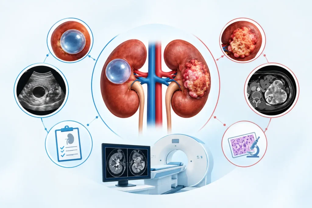
Symptoms alone rarely help distinguish a renal cyst vs kidney cancer, because both are often silent in their early stages. This is why imaging plays the central role in diagnosis, rather than physical exams or blood tests alone.
Doctors rely on a combination of scan type, contrast behavior, and structural features to make this distinction accurately and confidently.
How Doctors Use CT and Ultrasound to Tell Them Apart
Ultrasound is often the first imaging test used, since it is quick, radiation-free, and effective at identifying simple, fluid-filled cysts. However, when a mass looks even slightly complex on ultrasound, a contrast-enhanced CT scan is usually the next step.
On a CT scan, radiologists look closely at several key features:
- Wall thickness: thin and smooth suggests a cyst; thick or irregular raises concern.
- Presence of septations, or internal dividing walls, within the mass.
- Whether the mass enhances, meaning it absorbs contrast dye, a strong indicator of solid tissue and possible cancer.
- Calcification patterns within the cyst or mass wall.
- The overall shape and border of the mass, smooth versus irregular.
The Bosniak Classification: A Doctor’s Roadmap for Renal Cyst vs Kidney Cancer
To standardise this decision, radiologists and urologists use the Bosniak classification system, which grades kidney cysts from I to IV based on their imaging features and cancer risk.
| Bosniak Category | Imaging Features | Cancer Risk |
| I | Simple cyst, thin wall, no septa, no enhancement | Essentially none |
| II | A few thin septa, fine calcification, minimal enhancement | Very low |
| IIF | More septa or slightly thicker walls, needs follow-up imaging | Low, but monitored |
| III | Thickened irregular walls or septa with measurable enhancement | Roughly 40-60% |
| IV | Clearly solid, enhancing components within the mass | Over 85% |
Signs a Kidney Mass Could Be Cancerous
While most kidney masses are benign, certain imaging and clinical signs raise suspicion for cancer and prompt closer evaluation:
- A solid mass rather than a fluid-filled one.
- Irregular or thickened walls with measurable contrast enhancement.
- Rapid growth on repeat imaging compared to a prior scan.
- Blood in the urine, especially when combined with an imaging finding.
- Unexplained flank pain or a palpable mass on examination.
None of these signs alone confirms cancer, but together they guide a urologist toward recommending further testing or a biopsy.
When Is a Biopsy Needed to Confirm Renal Cyst vs Kidney Cancer?
Not every kidney mass needs a biopsy. Simple cysts, Bosniak I and II lesions, rarely require any tissue sampling because imaging alone is reliable enough.
A biopsy becomes useful mainly for indeterminate masses, where imaging features are unclear, or when the result would directly change the treatment plan, such as confirming cancer before surgery in a patient who prefers to avoid an operation until certain.
Renal Cyst vs Kidney Cancer: How Treatment Differs
Once doctors have classified a mass, treatment paths diverge significantly:
- Simple cysts (Bosniak I-II): usually require no treatment, just routine monitoring if at all.
- Bosniak IIF lesions: monitored with follow-up imaging at set intervals to watch for change.
- Bosniak III lesions: often surgically removed or closely evaluated given the meaningful cancer risk.
- Bosniak IV lesions and confirmed kidney cancer: typically treated with partial or complete nephrectomy, depending on tumor size and location.
This is exactly why an accurate distinction between a renal cyst vs kidney cancer at the imaging stage is so important, as it directly determines whether a patient needs surgery, monitoring, or nothing at all.
When to Consult a Kidney Cancer Specialist in Indore
If a scan report mentions an indeterminate or complex kidney mass, it is worth getting an urologist’s interpretation rather than relying on the radiology report alone. A specialist can correlate the Bosniak category with your symptoms, history, and overall risk to recommend the right next step.
Why Choose Dr. Vikas Singh for Kidney Cancer Evaluation in Indore
Dr. Vikas Singh (MBBS, MS, MCh) is widely regarded as the best kidney cancer doctor in Indore, offering careful imaging review and honest guidance on whether a kidney mass needs monitoring, biopsy, or surgery.
As an experienced kidney cancer specialist in Indore, he reviews each scan in the context of the Bosniak classification and the patient’s overall health, rather than reacting to a report in isolation. For patients searching for kidney cancer treatment in Indore, or simply an urologist doctor in Indore to make sense of a confusing scan finding, Dr. Vikas Singh, a trusted kidney cancer doctor in Indore, offers clear, judgment-free evaluation.
As a leading male urologist in Indore and widely regarded as the best urologist in Indore for complex kidney cases, he ensures every patient understands exactly what their scan means before deciding on next steps.
Book Your Kidney Mass Evaluation in Indore
| Found a kidney cyst or mass on your scan and unsure what it means? Get an expert second opinion before you worry unnecessarily or delay important care. Dr. Vikas Singh, regarded as the best kidney cancer doctor in Indore, offers accurate scan review and honest, clear guidance. Call +91 81468 73931 or book your appointment online today. |
Frequently Asked Questions
1. Can a kidney cyst turn into cancer?
A simple kidney cyst almost never turns into cancer. However, a complex cyst with irregular walls, septations, or contrast enhancement carries a higher risk and needs closer monitoring or further evaluation, which is why Bosniak classification matters.
2. How do doctors know if a kidney mass is cancerous on a scan?
Doctors look at features like wall thickness, internal septations, and whether the mass enhances with contrast dye on a CT scan. A solid, enhancing mass with irregular borders is far more likely to be cancerous than a simple, thin-walled fluid-filled cyst.
3. Is a renal cyst dangerous?
Most renal cysts are completely harmless and are often discovered by accident during an unrelated scan. They typically need no treatment, though larger or complex cysts may require monitoring or, rarely, drainage if they cause pain.